
Chest Radiograph of the 70 year old female presenting with large mass growth in the sternal region showing significant widening of the superior mediastinum.
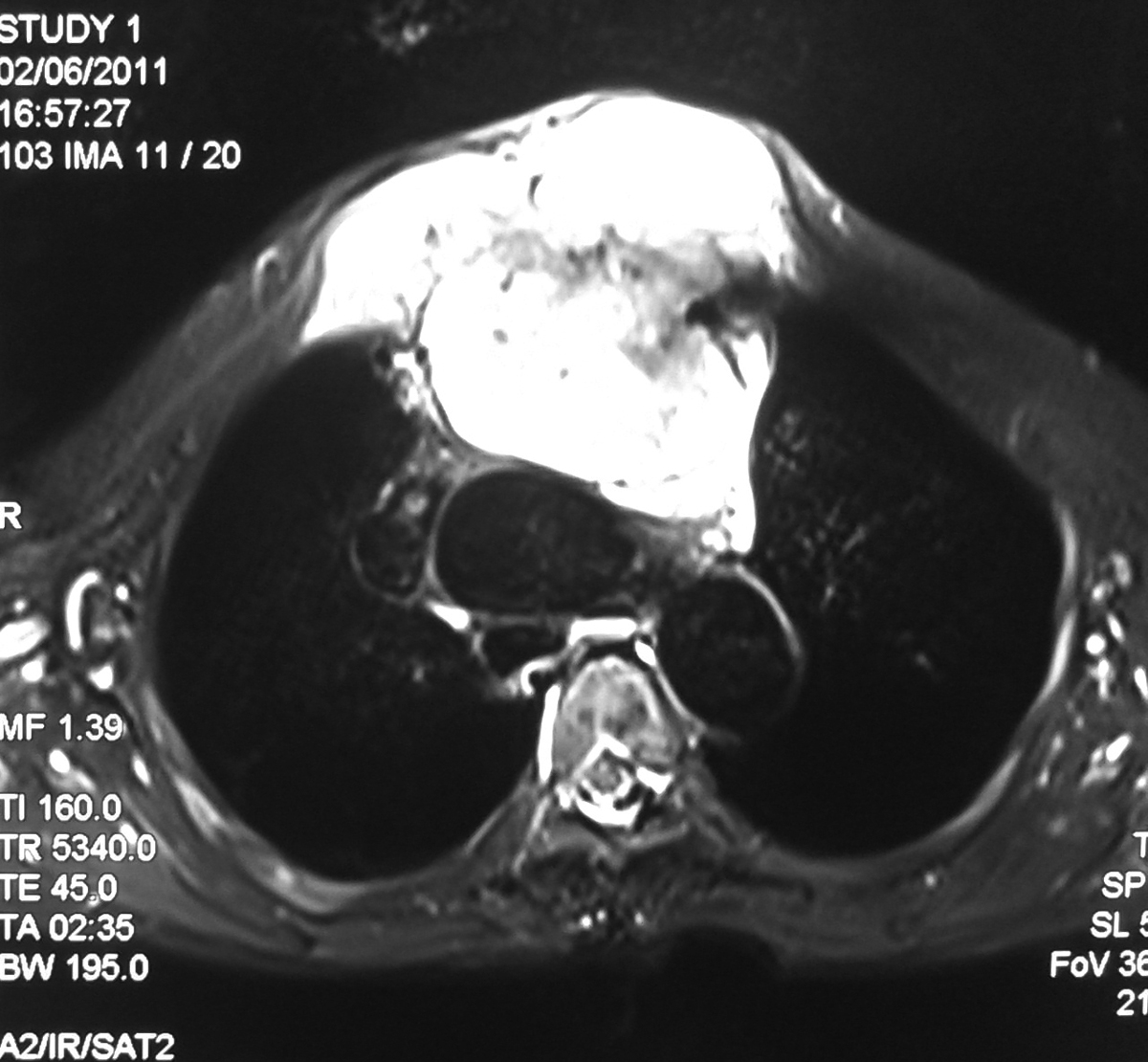
Axial T2 FS image showing large hyperintense mass lesion seen arising from the sternum with large retrosternal component causing mild mass effect on the mediastinal vessels.

T1 weighted axial image showing the lesion is hypo to isointense with retrosternal component.

Sagittal image showing large lesion in the sternal region and also extending superiorly in to the neck causing mass effect on the trachea.
Discussion:
- It represent ~ 30% of primary malignant bone neoplasias, being the most frequent that of the anterior thoracic wall.
- This tumor occurs more often between the third and fourth decade of life.
- The males are more affected
- They are lobulated neoplasias that may grow to massive proportion and, consequently, may extend internally to the pleural space, or externally, invading muscle and adipose tissue of the thoracic wall.
- Microscopically, the findings vary from normal cartilage to obvious malignant modifications. The differentiation between chondroma and chondrosarcoma can be extremely difficult
- Palpable mass in thorax is the main symptom in approximately 80% of the patients with thoracic wall tumor. Of these, 60% present associated pain
- Respiratory failure and hemothorax are rare, and are present only in very large tumors
- Imaging exams may be useful to indicate the pathology and extent of the lesion; however, the definitive diagnostic requires a correlation between histology and radiology.
- Computerized tomography (CT) and magnetic resonance (MR) are good exams to characterize the tumor and its extension.
- CT is superior to MR to demonstrate calcifications, whereas MR is the choice to evaluate the tumor’s extension and its relationships with adjacent structures
- Thoracic wall chondrosarcomas typically grow slowly and relapse locally. If not treated, late metastasis will occur.
- Complete control of the primary neoplasia is the main determinant of survival. The purpose of the first surgery must be a wide resection, enough to prevent local recurrence.

No comments:
Post a Comment