
CT axial section of chest lung window in 50 year old male showing diffuse bilateral symmetrical patchy ground glass opacities with areas of air space opacities, interlobular septal thickening and peribronchovascular thickening. Features consistent with Pneumocystis carinii pneumonia.

CT scan plain image axial section through superior aspect of liver showing few small simple cysts in segment VII.
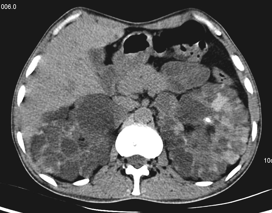
CT scan axial section at the level of kidneys showing enlarged bilateral kidney and they are replaced by multiple cystic lesions of variable sizes. Features consistent with Austosomal dominant polycystic kidney disease.


