Thursday, January 12, 2012

Elbow Dislocation

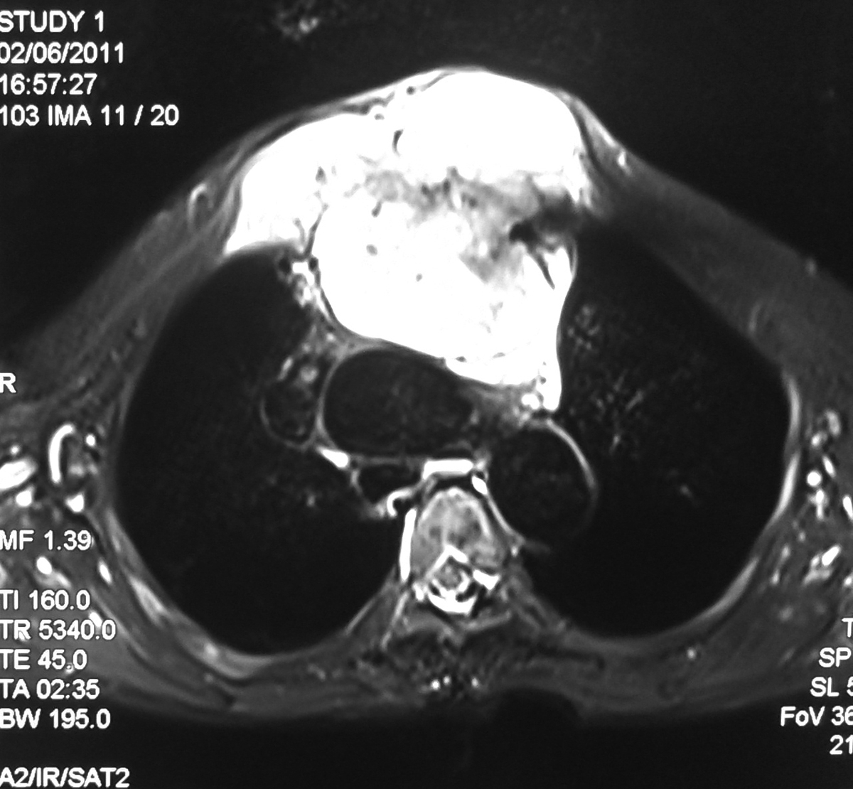
32 Year old male with road traffic accident showing posterior dislocation of the elbow joint with mild proximal migration of the radius and ulna and small displaced chip fracture of the coronoid process.

MRI sagittal T2 images showing posterior dislocated elbow.


Discussion:
  • Elbow dislocation is the second most common major joint dislocation.
  • Dislocation is usually closed and posterior.
Mechanism:
  • Dislocations of elbow usually result from fall onto extended elbow. 
  • Anatomic morphology of semilunar notch may predispose to elbow dislocation.
  • Central angle of semilunar notch is significantly larger in group of pts who had dislocation of the elbow compared to normals.
Classification:
  • Classified according to direction of dislocation, namely posterior, posterolateral, posteromedial, lateral, medial, or divergent.
  • Simple - elbow dislocations without fracture.
  • Complex - dislocations with fracture. 
Simple dislocation:
  • Rupture of capsule, rupture of MCL, lateral ligaments, rupture of flexor pronator mass and less commonly, injury to brachialis muscle.
  • Rupture of brachial artery has been reported.
Complex dislocation:
  • Dislocation with radial head fracture is most common complex dislocation as in our case.
  • Fracture dislocation with MCL injury: radial head fracture & MCL Instability: 
  • Terrible Triad: (dislocation, cornoid process #, and radial head #).
Vascular injury: Closed dislocation rarely associated with vascular injury.

Neurologic injury:
  • Compartement syndrome.
  • Neuropraxia.
  • Ulnar nerve injury in 14%.
  • Entrapment of median nerve.

Wednesday, January 11, 2012

Phenytoin associated Cerebellar atrophy - MRI


28 year old female with chronic seizures on long term phenytoin therapy. MR examination of the brain showing diffuse atrophy of the both lobes of the cerebellum, vermis and the brainstem evidenced by prominent cerebellar foliae, pre pontine and CP angle cisterns.
Discussion:
Diffuse atrophy of the cerebellum refers to degeneration / reduction from a previously normal cerebellar volume. The term slightly differs from cerebellar hypoplasia meaning the cerebellum was not well formed to start with.
  • Diffuse atrophy can result from variety of causes
  • Normal ageing
  • Alcoholic cerebellar degeneration
  • Drugs (e.g phenytoin)
  • Multi system atrophy (MSA)
  • Ataxia telangiectasia 
  • Friedreich ataxia
  • Marinesco-Sjögren syndrome (MSS)
  • Fahr disease : atrophy not specific to cerebellum
  • Paraneoplastic cerebellar degeneration (PCD)
  • Olivopontocerebellar atrophy (OPCA)
  • Cerebrotenidinous xanthomatosis

Differential diagnoses
  • Cerebellar hypoplasia
  • Mega cisterna magna

Chondrosarcoma of Sternum - MRI

Chest Radiograph of the 70 year old female presenting with large mass growth in the sternal region showing significant widening of the superior mediastinum.

Axial T2 FS image showing large hyperintense mass lesion seen arising from the sternum with large retrosternal component causing mild mass effect on the mediastinal vessels.

T1 weighted axial image showing the lesion is hypo to isointense with retrosternal component.

Sagittal image showing large lesion in the sternal region and also extending superiorly in to the neck causing mass effect on the trachea.

Discussion:
  • It represent ~ 30% of primary malignant bone neoplasias, being the most frequent that of the anterior thoracic wall.
  • This tumor occurs more often between the third and fourth decade of life.
  • The males are more affected 
  • They are lobulated neoplasias that may grow to massive proportion and, consequently, may extend internally to the pleural space, or externally, invading muscle and adipose tissue of the thoracic wall. 
  • Microscopically, the findings vary from normal cartilage to obvious malignant modifications. The differentiation between chondroma and chondrosarcoma can be extremely difficult 
  • Palpable mass in thorax is the main symptom in approximately 80% of the patients with thoracic wall tumor. Of these, 60% present associated pain 
  • Respiratory failure and hemothorax are rare, and are present only in very large tumors 
Imaging:
  • Imaging exams may be useful to indicate the pathology and extent of the lesion; however, the definitive diagnostic requires a correlation between histology and radiology. 
  • Computerized tomography (CT) and magnetic resonance (MR) are good exams to characterize the tumor and its extension. 
  • CT is superior to MR to demonstrate calcifications, whereas MR is the choice to evaluate the tumor’s extension and its relationships with adjacent structures 
  • Thoracic wall chondrosarcomas typically grow slowly and relapse locally. If not treated, late metastasis will occur. 
Treatment:
  • Complete control of the primary neoplasia is the main determinant of survival. The purpose of the first surgery must be a wide resection, enough to prevent local recurrence.

Saturday, January 7, 2012

Cysticercosis (Muscular)- USG

Ultrasonography showing a well defined thick walled anechoic lesion with posterior acoustic enhancement seen in the medial aspect of the distal arm within the biceps brachi muscle. The lesion shows echogenic mural nodule attached to the posterior wall.

Transverse view of the same lesion showing cystic lesion with mural nodule

 Colour Doppler showing no evidence of colour flow in the lesion

Power Doppler showing no flow in the lesion.
The findings are consistent with Cysticercosis. Confirmed on HPR.

Uterine Arteriovenous Malformation - USG Doppler

Gray scale trans abdominal longitudinal view Ultrasonography of uterus in a 34 year old female showing multiple an-echoic areas within the myometrum of the anterior wall (arrow) displacing the thin linier endometrium (arrow head).

On colour Doppler there are multiple colour filled dilated vascular channels.

Power Doppler depicting the color flow within the lesion

Spectral wave form at the feeding artery showing high velocity and relatively high resistant arterial flow.

Spectral tracing in the draining vein showing venous flow. The findings are consistent with uterine arteriovenous fistula. Patient had history of recent medical termination of pregnancy with dilatation of curettage.

Discussion:

Uterine vascular malformations include:
  • Arteriovenous malformations (AVM) (especially arteriovenous fistulas).
  • True aneurysms.
  • Pseudoaneurysms. 
Uterine AVMs are rare in nonpregnant women.
It is otherwise called with several terms which are cavernous hemangioma, cirsoid aneurysm, racemose aneurysm, arteriovenous aneurysm, pulsatile angioma, and arteriovenous fistula.

Etiology:
AV fistulas are usually acquired and typically represent a single artery joining a single vein.
They result as a consequence of previous uterine trauma (eg, prior pelvic surgery, curettage), use of intrauterine contraceptive devices, pathologic pregnancy-related events, and previous treatment for gestational trophoblastic disease.

Clinical Features:
Vaginal bleeding- Most common.
Congestive heart failure - secondary to steal phenomenon - Less common.
Imaging findings:
USG:
  • Gray-scale US shows a normal-appearing endometrium, whereas the myometrium contains multiple hypoechoic or anechoic spaces.
  • At color Doppler US, these cystic spaces generate color signals in a mosaic pattern representing turbulent flow.
  • Spectral analysis of the arterial vessels within the lesion shows high-velocity flow with a low resistive index (approximately 0.51–0.65). Spectral analysis of venous flow demonstrates a similar pattern.
CT: CT angiography of the pelvic vessels may reveal the vascular malformation.

MRI:
Multiple dilated tubular channels showing flow related signal voids (hypointense) on T1 and T2 images seen in myometrium with normal endometrium or displaced endometrium. The flow relted signal void channels may or may not extend in to the parametrium.

Treatment:
  • Acute treatment - Hemodynamic stabilization and management of active bleeding. 
  • Occlusion with a Foley catheter bulb may be effective. 
  • If fertility is not an issue, hysterectomy is the treatment of choice 
  • They may be treated successfully with intra-arterial embolization

*******

Search This Blog