
Coronal CT paranasal sinuses showing well defined rounded hyperdense focus seen in the right ethmoid air cell suggestive of ivory osteoma
All the sinuses shows soft tissue opacification including the nasal cavity due to allergic fungal rhinosinusitis.
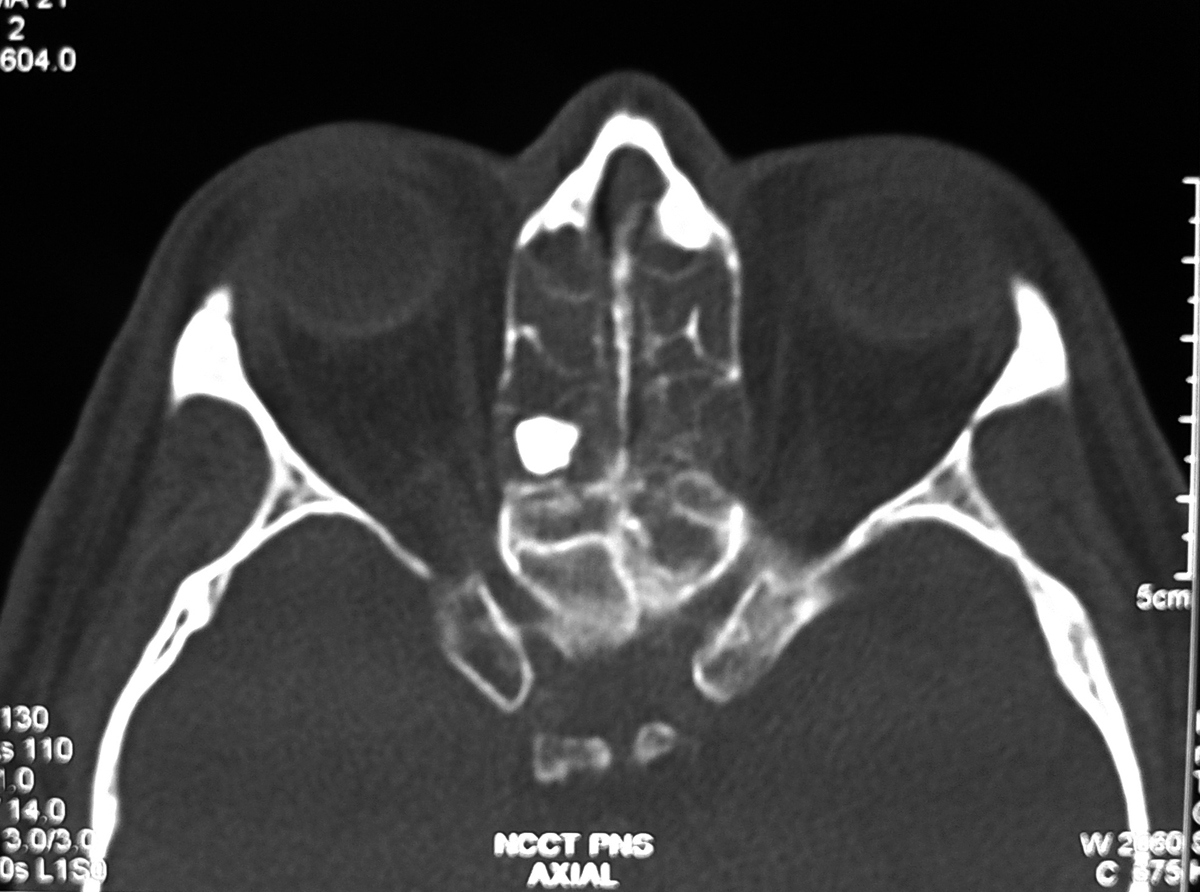
Axial CT section showing the Ivory osteoma in the posterior ethmoid air cell
Discussion:
Most common tumor of the paranasal sinuses and frequently seen in the frontal and ethmoid sinuses. It is benign tumor of membranous bone consisting of dense, compact bone and majority of of them are discovered serendipitously.In the skull, they usually arise from the outer table. Rarely, large osteoma in the frontal or ethmoid region may displace globe forward and cause proptosis.
Obstruction of a sinus ostium may lead to infection or formation of a mucocele. Very rarely, an osteoma may erode through the dura leading to cerebrospinal fluid rhinorrhea or intracranial infection.
Imaging findings:
Well-circumscribed, sharply-marginated round and very dense lesions usually less than 2 cm in size. Multiple paranasal osteomas are found in Gardner’s syndrome.
Multiple osteoma of the mandible and maxilla, along with the frontal, sphenoid and ethmoid sinuses, rarely the long bones or phalanges
Association between colonic polyps with a predilection to malignant degeneration.
Multiple osteoma of the mandible and maxilla, along with the frontal, sphenoid and ethmoid sinuses, rarely the long bones or phalanges
Association between colonic polyps with a predilection to malignant degeneration.

No comments:
Post a Comment