
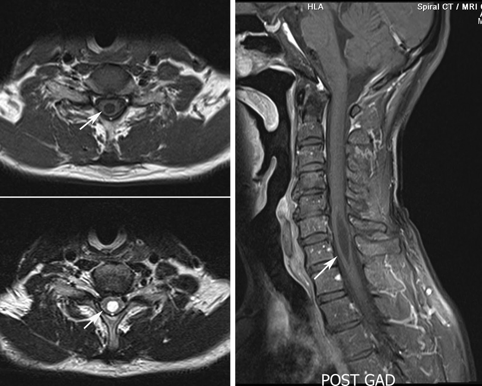
Cervical spine MRI in a 54 year old male patient showing focal dilatation of the central spinal canal with no enhancing focal lesion within the cord. The features are consistent with the focal syrinx.
Discussion:
Hydromyelia: An accumulation of cerebrospinal fluid (CSF) may lead to simple distention of the central canal of the spinal cord lined by ependymal cells.
Syringomyelia: an accumulation of CSF may dissect into the surrounding white matter to form a paracentral cavity, which is not lined by ependyma.
syringohydromyelia: Combination of both which is seen in most of the cases.
I - In 1973, Barnett et al classified syringohydromyelic cavities into 5 types:
- Communicating (with the subarachnoid space, usually at the level of the obex at the inferior aspect of the fourth ventricle)
- Posttraumatic
- Tumor-related
- Arachnoiditis-related
- Idiopathic.
II - Milhorat et al, the intramedullary cavities were classified into communicating, noncommunicating, and atrophic types.
III - Noncommunicating syringes are subdivided into 6 types:
- Chiari II malformation with hydrocephalus
- Chiari I malformation without hydrocephalus
- Extramedullary compressive lesions at the craniocervical junction or along the length of the spinal canal
- Spinal cord trauma
- Intramedullary tumors and intraperimedullary infections
- Multiple sclerosis

No comments:
Post a Comment