Sunday, September 11, 2011

Usual Interstitial Pneumonia - HRCT

HRCT of 40 Year old female showing macrocystic honeycombing, reticular
opacities predominantly in the subpleural region, architectural distortion and
areas of focal ground-glass opacity 

Mediastinal window of the same patient showing small enlarged pre tracheal lymphnode.

Discussion:

Idiopathic Pulmonary Fibrosis (IPF) is the most common entity of the Idiopathic Interstitial Pneumonias (IIPs). By definition, IPF is the term for the clinical syndrome associated with the morphologic pattern of UIP.
Median survival - 2 to 4 years.

Clinical features - Presents in 50 years old or older. Patients present with progressively worsening
dyspnea and nonproductive cough.

Histopathologic Features: Spatial and temporal heterogeneity, architectural distortion and fibroblastic foci.

HRCT: Distribution is apicobasal gradient and predomonently subpleural location. 
Features are macrocystic honeycombing, reticular opacities, traction bronchiectasis, architectural distortion and focal ground-glass opacity.

Saturday, September 10, 2011

Temporal bone fracture with facial nerve injury - HRCT

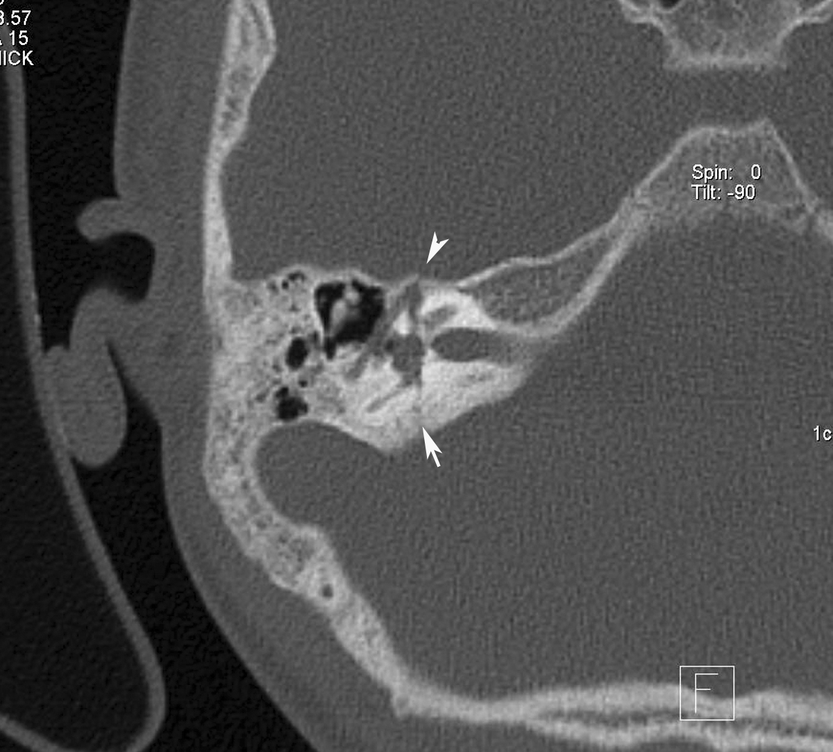
HRCT of right temporal bone axial image showing transverse temporal bone fracture (arrow) and the fracture line passing through the first anterior genu of the facial nerve canal (arrow head).

Subsequent section of the HRCT right temporal bone showing the fracture passing through the facial nerve canal (arrow head).

Discussion: Temporal bone fractures classically are described with reference to the long axis of the petrous bone.
Classification:
1. Longitudinal (parallel to the axis).
2. Transverse (perpendicular to the axis).
3. Oblique/Mixed- Common.


Longitudinal fracture: The fracture line of force runs roughly from lateral to medial. A fracture line may extend through the facial nerve canal, thereby damaging the facial nerve. Associated injury, such as transection or intraneural hemorrhage, may cause facial nerve paralysis, as can damage from displaced bone fragments.


Transverse fracture: result from trauma to the occiput or cranial-cervical junction, with the line of force running roughly anterior to posterior.A fracture passing through the vestibulocochlear apparatus can cause sensorineural hearing loss and equilibrium disorders. Transverse fractures also commonly injure CN VII, because their path often takes them close to the nerve's labyrinthine segment.

Mixed fractures: Oblique (or mixed) fracture patterns, which extend both longitudinally and transversely, are common, and some case series report that these occur more often than do isolated transverse or longitudinal fractures.

Radiology:
Radiography: Plain film radiographs of the skull may show opacified mastoid air cells, intracranial air, or, rarely, a lucency (fracture line).

HRCT: can demonstrate a lucency through the temporal bone. Involvement of the middle ear, petrous bone, otic capsule, and facial nerve canal are the primary determinants of prognosis.


MRI: may demonstrate fluid (high signal on T2-weighted images) in the middle ear and mastoid air cells. T1-weighted images may reveal a bright signal in the labyrinth or middle ear, consistent with hemorrhage.

Splenic Artery Aneurysm in Extrahepatic Portal Vein Obstruction - CT Angio

Axial thick MIP image of CT angiography showing aneurysm arising from the main splenic artery

Coronal thick MIP image of CT angiography showing aneurysm arising from the main splenic artery

3 D volume rendering image of CT angiography showing aneurysm arising from the main splenic artery (arrow).

Axial CT image in venous phase showing multiple collateral veins in the porta with non visualised main portal vein consistent with extra heaptic portal vein obstruction. Note splenomegaly and multiple leinorenal collaterals.

Discussion:

Splenic artery aneurysms (SAA) are the most common visceral aneurysm occuring predominantly in females. SAA causing EHPVO and presenting with features of portal hypertension is extremely rare.
Women are affected four times more commonly by SAA and often during pregnancy and childbearing years due to endocrine changes.
The usual location of splenic artery aneurysm is at the mid or distal portion of the splenic artery, frequently found at the arterial bifurcation.

Clinical features: They are usually asymptomatic, and the symptomatic presentation includes chronic abdominal pain of varied severity or an acute rupture with hypotension.

Radiology:
CT angiography is the best modality for the diagnosis, although it is usually detected incidentally.

Treatment:
Endovascular: Coiling or covered stent.
Operative: Aneursyectomy with or without splenectomy.

Friday, September 9, 2011

Tracheal Foreign Body - CT Scan with 3D.

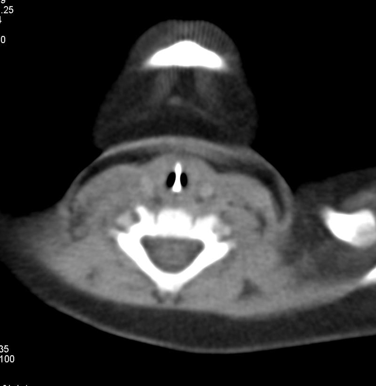
CT scan axial soft tissue window of 7 Months old male child with history of foreign body aspiration with asymptomatic baby showing linier metallic foreign body oriented vertically in the center with patent lumen on either side. 

Coronal reformatted CT scan soft tissue window well delineate the vertical extent of the foreign body

Minimum intensity projection (minIP) image showing the foreign body. 

3D Volume rendered image showing showing the foreign body. 

Virtual bronchoscopy showing showing the foreign body (arrow). 

Extraction by bronchoscopy is the treatment of choice for tracheal foreign bodies

Tuesday, August 23, 2011

Gray matter heterotopia - MRI


T1, T2 and T1 inversion recovery axial image in 16 year old female patient with seizures showing smooth, nodular area, isointense to grey matter seen on all the sequences and present all along the margin of the bilateral lateral ventricles (subependymal regions) consistent with gray matter heterotopia.The lesion is better depicted in T1 inversion recvoery image.

Discussion:
Definition: It is a type of cortical dysplasia (neuronal migrational anomaly) where in clumps of normal grey matter being located in the wrong part of the brain.
Clinical features: Asymptomatic to profound seizures and it is usually detected incidentally on brain MRI. Various manifestations include severe seizure disorder, loss of motor skills, and mental retardation. Fatalities are practically unknown, other than the death of unborn male fetuses with a specific genetic defect.
Types of Heterotopia:
  1. Periventricular or subependymal: It can be seen as small single node or a large number of nodes, can exist on either or both sides of the brain at any point along the higher ventricle margins, can be small or large, single or multiple, and can form a small node or a large wavy or curved mass
  2. Focal Subcortical: form as distinct nodes in the white matter, "focal" indicating specific area.
  3. Band Form: "Band" heterotopia form in the white matter beneath the cortex, but the gray matter is more diffuse and is symmetric between the hemispheres.
Imaging: CT or MRI
CT scan: The density will be similar to that of the gray matter.
MRI: The signal intensity will be similar to gray matter in all the sequences. T1 inversion recovery sequence will better depict the lesion.

Thursday, July 7, 2011

Hemangioendothelioma-CT


3 month old male child came with clinically enlarged nodular palpable liver. CT scan shows enlarged liver with multiple peripherally enhancing variable sized nodular lesion seen occupying whole of the liver in both the lobes. Infra renal part of aorta is small in caliber (arrow head) with dilated and prominant celiac axis (not shown here). There is also similar lesion seen in the upper back subcutaneous region on right side. Features suggestive of hemangioendothelioma.

Discussion:

Benign hemangioendothelioma is a rare liver neoplasm, however it represents the most common vascular tumor of the liver in the neonate. 
The neonatal clinical presentation of hemangioendothelioma includes: 
1) gross liver enlargement, 
2) high-output cardiac failure, and 
3) associated hemangiomas in other organs including skin (hemangiomatosis).

Other complications include consumptive coagulopathy, hemolytic anemia, tumor rupture, and problematic surgical intervention. Hemangioendotheliomas have been recognized on obstetric ultrasound. Neonatal CT, repeat sonography, MRI imaging, and arteriography have all been utilized to visualize the tumor and assess it's size, location, and the anatomy of arterio-venous malformation (AVM) in the neonate. 

The antenatal ultrasound features of hemangioendothelioma include: liver mass (heterogeneous, hypoechoic, complex with anechoic spaces, and hyperechoic), consequent liver enlargement, prominent vasculature, cardiomegaly, and possible hydrops (serous effusions and body edema).Color and pulsed Doppler interrogation can also be particularly useful in identifying an AVM.Post natal findings are liver enlargement, multiple or single confluent liver masses showing peripheral enhancement, may find AV malformation. Typically the celiac axis will be dilated and reduced caliber of infra renal aorta. May find extrahepatic hemangioendothelioma.

Monday, July 4, 2011

Neurocysticercosis starry sky appearance - MRI

Contrast enhanced T1 weighted axial image showing multiple nodular and ring enhancing lesions in bilateral cerebral hemispheres giving starry sky appearance.

Contrast enhanced T1 weighted coronal image showing multiple nodular and ring enhancing lesions in bilateral cerebral hemispheres.

Colloid cyst - MRI

Axial non enhanced T1 magnetic resonance image showing a bright mass in the roof of third ventricle (foramen of Monro).

Axial non enhanced T2 magnetic resonance image showing a hypointense mass in the roof of third ventricle.

Sagittal non enhanced T1-weighted magnetic resonance image. This image demonstrates a round area of increased signal intensity lesion in the anterosuperior portion of the third ventricle.

Discussion:
Colloid cysts are benign, congenital epithelium-lined cysts that almost always arise in the anterior third entricle. However, rare reports describe cysts in other locations. The cysts are believed to derive from either primitive neuroepithelium of the tela choroidea or from endoderm.
Patient may present with intermittent head ache. Rarely m
ay cause sudden death .

CT scan:
  • Rounded small mass with high attenuation at the anterior third ventricle - due to proteinacious material.
  • Rarely the lesion is isoattenuating or hypoattenuating relative to brain parenchyma.
  • Intermittent hydrocephalus - colloid cyst act as ball valve.
MRI:
  • Usually found at the foramen of Monro just posterior to the fornices in the anterosuperior third ventricle.
  • MR signal intensities are variable: Variation due to contents of the cyst.
  • Commonly hyperintense on T1 and iso to hypointense on T2.
  • Colloid cysts do not have intrinsic enhancement; the presence of enhancement suggests a solid tumor. Peripheral enhancement may be present because of vascularity in the outer wall.

Traumatic AVN of Scaphoid

PA radiograph of the wrist with scaphoid deviation shows a fracture of the waist of the scaphoid bone (arrow) with loss of trabecular pattern in the distal fragment.

T1 weighted images of the same patient 3 months after trauma showing hypointensity in the distal fractured fragment consistent with avascular necrosis.

Discussion:
Scaphoid fractures are the most common carpal fractures, resulting from a fall on an outstretched hand. 70 % of these occur at the waist, 20 % at the proximal pole, and 10 % at the distal pole. Blood supply for the proximal pole enters at the waist. If this blood supply is interrupted due to fracture, the proximal pole is at risk for avascular necrosis unlike out case where the distal fragment is involved. Special scaphoid views with the hand in ulnar deviation may be needed to detect these fractures.

Avascular necrosis
  • More common in scaphoid because of peculiar blood supply
  • Up to 30% of scaphoid fractures may display increased density of the proximal pole
  • Often reversible
  • May be due to relative ischemia of proximal pole
  • Occurs in 15-30% of scaphoid fractures
  • Almost always involves proximal pole
  • The more proximal is the fracture line, the risks of avascular necrosis increase
  • The radiographic hallmarks of AVN are collapse and fragmentation
  • MRI may be more sensitive to AVN than conventional radiographs but is not 100% sensitive.

Sunday, July 3, 2011

Ivory Osteoma

Coronal CT paranasal sinuses showing well defined rounded hyperdense focus seen in the right ethmoid air cell suggestive of ivory osteoma
All the sinuses shows soft tissue opacification including the nasal cavity due to allergic fungal rhinosinusitis.

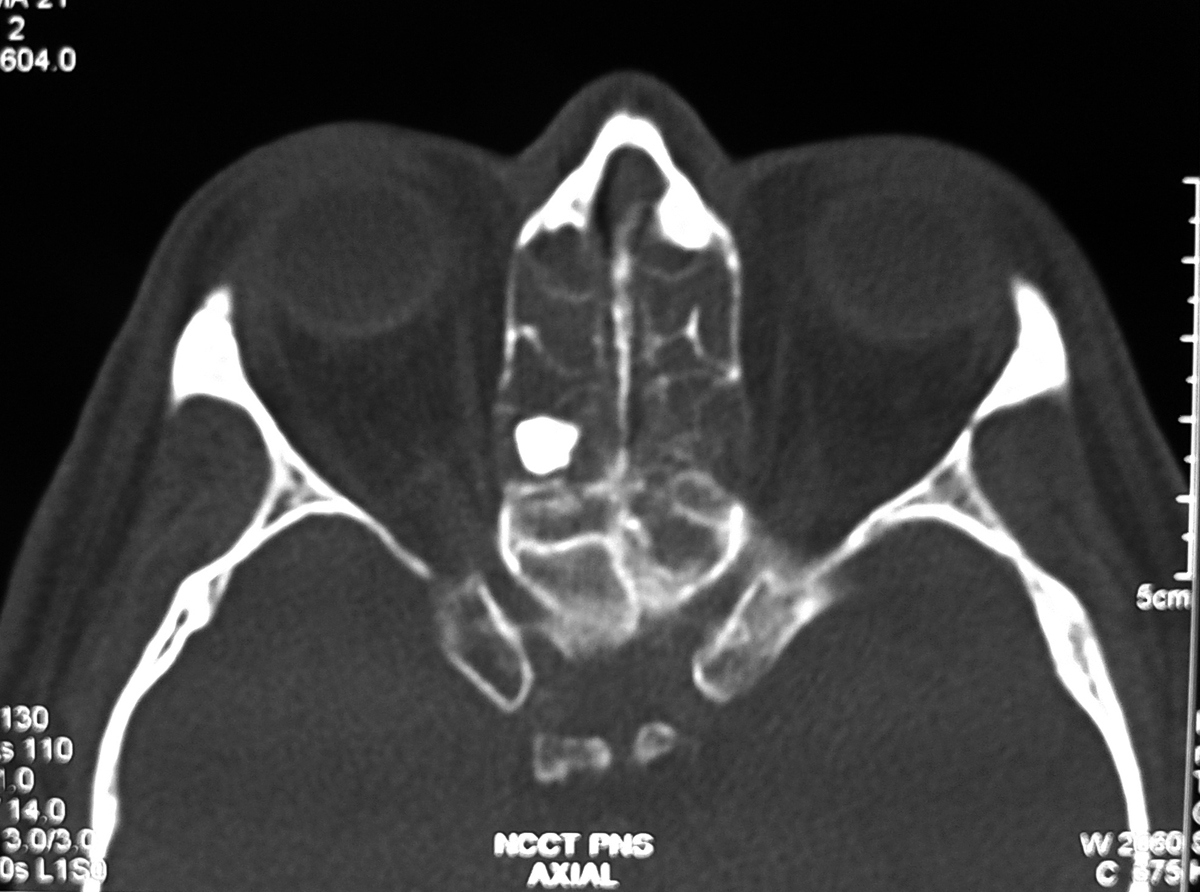
Axial CT section showing the Ivory osteoma in the posterior ethmoid air cell

Discussion:
Most common tumor of the paranasal sinuses and frequently seen in the frontal and ethmoid sinuses. It is benign tumor of membranous bone consisting of dense, compact bone and majority of of them are discovered serendipitously.
In the skull, they usually arise from the outer table. Rarely, large osteoma in the frontal or ethmoid region may displace globe forward and cause proptosis.
Obstruction of a sinus ostium may lead to infection or formation of a mucocele. Very rarely, an osteoma may erode through the dura leading to cerebrospinal fluid rhinorrhea or intracranial infection.

Imaging findings:
Well-circumscribed, sharply-marginated round and very dense lesions usually less than 2 cm in size. Multiple paranasal osteomas are found in Gardner’s syndrome.
Multiple osteoma of the mandible and maxilla, along with the frontal, sphenoid and ethmoid sinuses, rarely the long bones or phalanges
Association between colonic polyps with a predilection to malignant degeneration.

Friday, July 1, 2011

Traumatic fracture of terminal phalynx


Traumatic fracture in the base of the terminal phalynx (arrow).

Healed Varicella Pneumonia


There are multiple tiny subcentimeter calcific densities noted throughout both lung parenchyma more on the left side. There is no focal lung parenchymal mass or cavitating lesion seen. The appearances are classical for previous varicella exposure. 

Reference: 
Kim EA, et al. Viral Pneumonias in Adults: Radiologic and Pathologic Findings. RadioGraphics 2002; 22: 137.Credit: Dr Abhijit Datir

Lateral epicondyle bursitis

Coronal STIR image shows focal hyperintense signal (fluid signal) in the region of lateral epicondylar bursa suggestive of bursitis. No evidence of altered marrow signal intensities.

 Axial T2 fat suppressed image showing fluid collection in bursa.
Sagittal image showing the hyperintensities.

Discussion:
The inflammation of the lateral epicondyle bursa which should not be mistaken for the lateral epicondylitis/tennis elbow.
Lateral epicondylitis, colloquially refereed to as “tennis elbow“ presents as pain and tenderness over the common extensor tendon insertion on the lateral epicondyle elicited by active supination of the forearm and dorsiflexion of the wrist.
The MRI appearance of lateral epicondylitis is thickening and/or increased signal intensity in the common extensor tendon can be seen on T2-weighted spin echo, T2*-weighted gradient-echo or STIR images in the coronal plane. Fat-suppressed Fast spin-echo sequences are particularly useful. The underlying bone marrow may show changes.
The MRI appearance of the lateral epicondyle bursitis is variable fluid collection within the bursa. No marrow altered signal intensity seen.


Diaphyseal Aclasis-Hereditary multiple exostoses

Radiograph of left knee AP and lateral views showing multiple bony out growth seen in the metadiaphyseal region growing away from the epiphysis in femur, tibia and fibula.
Radiograph of another patient of bilateral knee AP views showing multiple bony out growth in the metadiaphyseal region growing away from the epiphysis in femur, tibia and fibula.

Discussion: Diaphyseal Aclasis is also known as:
1. External Chondromatosis Syndrome
2. Multiple Cartilaginous Exostoses
3. Multiple Exostoses
4. Multiple Exostoses Syndrome
5. Multiple Osteochondromatosis

Diaphyseal Aclasis isa relatively rare abnormal condition that affects the skeletal system. Characterized by multiple exostoses or bony protrusions, it is inherited as a dominant trait. Approximately half of the children of an individual with diaphyseal aclasis display varying degrees of its symptoms.Evident involvement is diffuse, with the long bones usually affected more severely and more frequently than the short bones. The malignant change is more frequent compared to the solitary exostosis.
Signs of malignant development refer my other post on Chondrosarcoma in osteochondroma -

Lipoma of the Filum Terminale

T1 sagittal image of the lumbar spine showing multiple osteoporotic collapsed vertebral bodies with elongated linier  wavy hyperintense intraspinal structure from the level of L2 to S1 consistent with filum terminale lipoma.

Axial T1 weighted image showing filum terminale lipoma as small intraspinal hyperintense dot (arrow).

Discussion:

Fat in the filum terminale is not an infrequent occurence, seen in 4 – 6%, and is usually easily detected on MRI (especially T1 sequences). It is usually an incidental finding of no clinical concern.
In some individuals however it is associated with spinal dysraphism, thickening of the filum terminale (>3mm) and tethering of the spinal cord.

Classification: Four patterns: 
1) fatty filum with descended conus medullaris and symptoms.
2) fatty filum with descended conus medullaris and no symptoms.
3) fatty filum with normal conus medullaris position and symptoms.
4) fatty filum with normal conus medullaris position and no symptoms

Imaging findings:
CT scan: If the lesion is large, seen as hypodense focus (fat density HU -90 to -30) below the level of conus. If it is  small may not be visible.

MRI: is modality of choice.
  • Hyperintense on T1 and T2 FSE
  • Saturates on fat saturation sequences
  • Demonstrates chemical shift artefact
  • No enhancement.
References
1. E Brown et al “Prevalence of incidental intraspinal lipoma of the lumbosacral spine as determined by MRI.” Spine. 1994 Apr 1;19(7):833-6.
2. K Koeller et al “Neoplasms of the Spinal Cord and Filum Terminale: Radiologic-Pathologic Correlation” RadioGraphics 2000; 20: 1721-1749.

Search This Blog