Showing posts with label Musculoskeletal radiology. Show all posts
Wednesday, August 1, 2012
Thursday, January 12, 2012
Elbow Dislocation
32 Year old male with road traffic accident showing posterior dislocation of the elbow joint with mild proximal migration of the radius and ulna and small displaced chip fracture of the coronoid process.
MRI sagittal T2 images showing posterior dislocated elbow.
Discussion:
- Elbow dislocation is the second most common major joint dislocation.
- Dislocation is usually closed and posterior.
- Dislocations of elbow usually result from fall onto extended elbow.
- Anatomic morphology of semilunar notch may predispose to elbow dislocation.
- Central angle of semilunar notch is significantly larger in group of pts who had dislocation of the elbow compared to normals.
- Classified according to direction of dislocation, namely posterior, posterolateral, posteromedial, lateral, medial, or divergent.
- Simple - elbow dislocations without fracture.
- Complex - dislocations with fracture.
- Rupture of capsule, rupture of MCL, lateral ligaments, rupture of flexor pronator mass and less commonly, injury to brachialis muscle.
- Rupture of brachial artery has been reported.
- Dislocation with radial head fracture is most common complex dislocation as in our case.
- Fracture dislocation with MCL injury: radial head fracture & MCL Instability:
- Terrible Triad: (dislocation, cornoid process #, and radial head #).
Neurologic injury:
- Compartement syndrome.
- Neuropraxia.
- Ulnar nerve injury in 14%.
- Entrapment of median nerve.
Wednesday, January 11, 2012
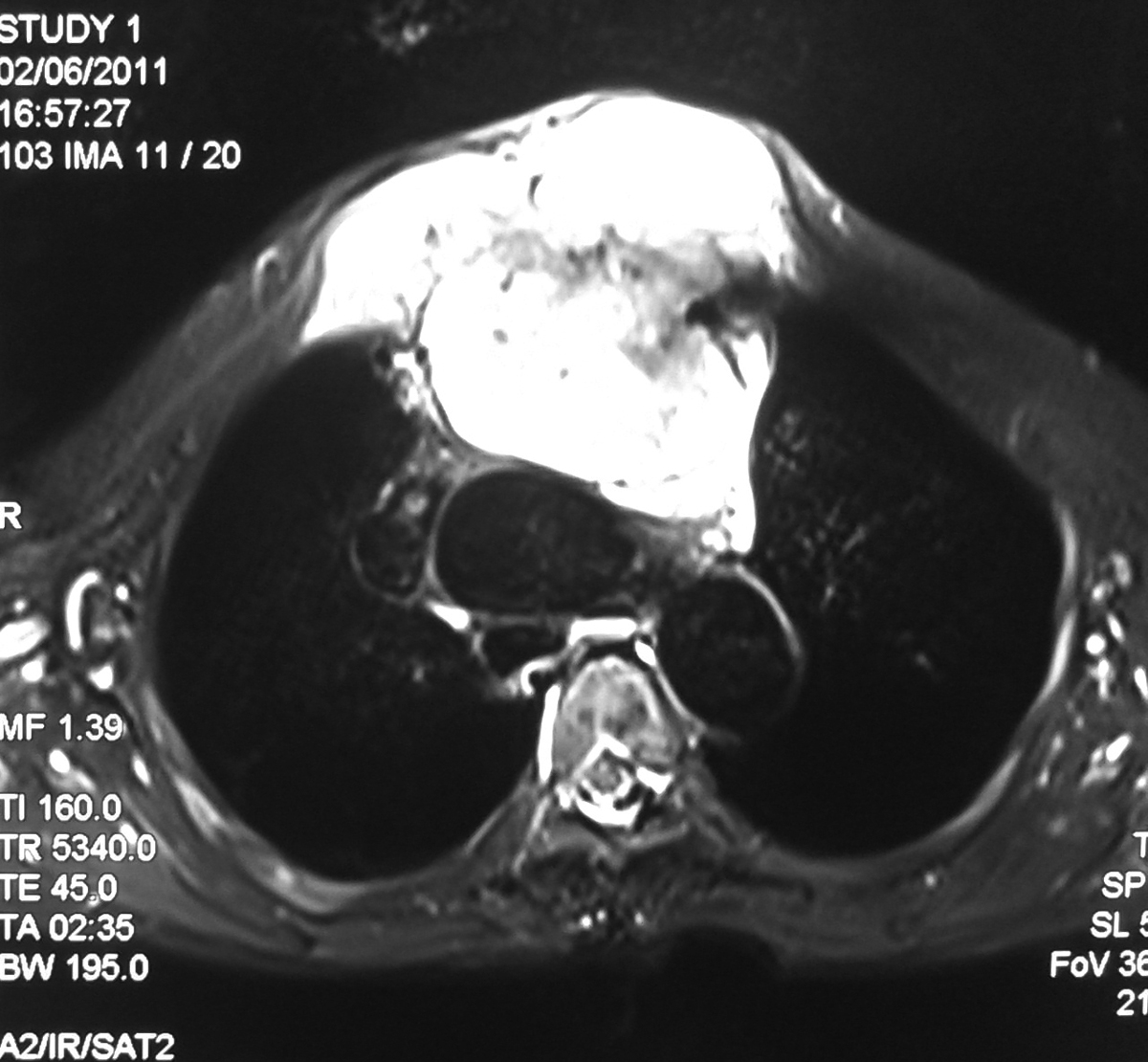
Chondrosarcoma of Sternum - MRI
Chest Radiograph of the 70 year old female presenting with large mass growth in the sternal region showing significant widening of the superior mediastinum.
Axial T2 FS image showing large hyperintense mass lesion seen arising from the sternum with large retrosternal component causing mild mass effect on the mediastinal vessels.
T1 weighted axial image showing the lesion is hypo to isointense with retrosternal component.
Sagittal image showing large lesion in the sternal region and also extending superiorly in to the neck causing mass effect on the trachea.
Discussion:
- It represent ~ 30% of primary malignant bone neoplasias, being the most frequent that of the anterior thoracic wall.
- This tumor occurs more often between the third and fourth decade of life.
- The males are more affected
- They are lobulated neoplasias that may grow to massive proportion and, consequently, may extend internally to the pleural space, or externally, invading muscle and adipose tissue of the thoracic wall.
- Microscopically, the findings vary from normal cartilage to obvious malignant modifications. The differentiation between chondroma and chondrosarcoma can be extremely difficult
- Palpable mass in thorax is the main symptom in approximately 80% of the patients with thoracic wall tumor. Of these, 60% present associated pain
- Respiratory failure and hemothorax are rare, and are present only in very large tumors
- Imaging exams may be useful to indicate the pathology and extent of the lesion; however, the definitive diagnostic requires a correlation between histology and radiology.
- Computerized tomography (CT) and magnetic resonance (MR) are good exams to characterize the tumor and its extension.
- CT is superior to MR to demonstrate calcifications, whereas MR is the choice to evaluate the tumor’s extension and its relationships with adjacent structures
- Thoracic wall chondrosarcomas typically grow slowly and relapse locally. If not treated, late metastasis will occur.
- Complete control of the primary neoplasia is the main determinant of survival. The purpose of the first surgery must be a wide resection, enough to prevent local recurrence.
Saturday, January 7, 2012
Cysticercosis (Muscular)- USG
Ultrasonography showing a well defined thick walled anechoic lesion with posterior acoustic enhancement seen in the medial aspect of the distal arm within the biceps brachi muscle. The lesion shows echogenic mural nodule attached to the posterior wall.
Transverse view of the same lesion showing cystic lesion with mural nodule
Colour Doppler showing no evidence of colour flow in the lesion
Power Doppler showing no flow in the lesion.
The findings are consistent with Cysticercosis. Confirmed on HPR.
Sunday, October 9, 2011
Lipoma Arborescens of the Knee - MRI
Sagittal T1 weighted MRI in 27 year old male shows a large suprapatellar effusion with a frond-like synovial mass of fat intensity in suprapatellar region and in posterior intercondylar region (arrows).
Sagittal PD FS MRI shows the synovial mass to be the same low intensity as fat (arrows).
Axial gradient echo MRI shows the characteristic frond-like pattern.
Discussion:
Lipoma arborescens is a rare benign lesion, which arises in the synovium and is characterized by villous proliferation of fat cells. The proliferating cells appear organic and often resemble a tree thus its name, arborescens, from the Latin arbor for tree.
Cause: is uncertain. One hypothesis is - synovial hyper-proliferation is in response to traumatic or inflammatory stimuli.
Associations: Trauma, osteoarthritis or rheumatoid arthritis.Radiograph: Findings are non specific.
1. Soft tissue density with areas of lucency suggesting fat.
2. Underlying degenerative changes of the knee.
MRI: is the investigation of choice.
- Villous proliferation, often with a frond-like configuration, which arises from within the synovial cavity.
- Fatty characteristics and typical pattern of proliferation shows isointense to the fat on all the sequences.
- Joint effusion.
- Meniscal tears.
- Baker's cyst in 38%.
Thursday, October 6, 2011
Scapholunate ligament tear - MRI
Radiograph of wrist in 45 Year old male patient with pain in the wrist joint showing widened scapholunate joint space with focal periosteal elevation in the lunate (arrow).
Coronal PD FS image showing complete tear in the scapholunate ligament with mild marrow edema in the scaphoid and lunate bones.
Coronal GRE image well depicts the ligament tear (arrow).
Discussion:- Most common and most significant ligament injury of wrist causing carpal instability.
- Risk factors: ulna minus configuation, slope of radial articular surface, and lunotriquetral coalition.
1. Dynamic scapholunate instability
- No radiographic evidence of malalignment is present (ie dynamic deformity);
- Diagnosis is established by dorsal S-L tenderness and positive shift test;
3. Scapholunate dissociation (SLD):
- SL ligament tear may lead to rotational dislocation of scaphoid allowing proximal pole to displace posteriorly & distal pole to displace anteriorly.
- Scaphoid inherently tends to palmar flex because of its oblique position and the loading applied through (STT) joint.
- Because scaphoid lacks proximal of ligament, it will rotate around radiocaptitate ligament leading to dorsal rotary subluxation of the proximal pole.
5. Scapholunate advanced collapse:
Osteosarcoma of femur - Radiograph and MRI
Radiograph of 20 year old female showing destructive lytic lesion involving the metaphysis of the medial aspect of femur with new bone formation and periosteal reaction (caudman triangle) {Arrow}
T1 weighted coronal image showing lytic destructive lesion with soft tissue, periosteal reaction and new bone formation.
Axial GRE image showing the new bone formation as hypointense signal and periosteal reaction as hyperintense signals.
Coronal STIR image showing the lesion.
Discussion: Osteosacroma is malignant tumor of bone in which neoplastic osteoid is produced by a proliferating spindle cell stroma.- Most common primary malignant bone tumor of mesenchymal derivation.
- Arises in adolescents, and second or third decade.
- Affects males slightly more often than females.
Affected sites:
- Occurs in region of knee (distal femur or proximal tibia) in 50% of patients.
- Other sites include proximal humerus, proximal femur, & pelvis.
- Most osteosarcomas occur in the metaphysis.
- Infrequently occurs in the spine.
- Classic
- Telangiectatic
- Parosteal
- Periosteal
Radiograph:
- Destructive lytic lesion or mixture of lytic and sclerotic areas (common).
- Moth eaten appearance with ill-defined zone of demarcation.
- Involves the metaphysis
- New bone formation.
- Periosteal reaction
- Visible soft-tissue mass.
- Spiculated / Sunburst appearance.
- 'Codman's Triangle' which is basically a subperiosteal lesion formed when the periosteum is raised due to the tumor.
CT Scan:
- Gives clearer indication of bone destruction.
- May depict small amounts of mineralized osseous matrix not seen on radiographs.
- May be particularly helpful in visualizing flat bones, in which periosteal changes may be more difficult to appreciate.
- Modality of choice in evaluating the local extent of disease because of its excellent bone marrow and soft tissue contrast and multiplanar capabilities.
- it assists in determining the most appropriate surgical management.
- Better delineates the involvement of the epiphysis, adjacent joint, adjacent soft tissues, neurovascular bundle, marrow extent and skip lesions.
- Increased uptake of radioisotope on bone scans obtained by use of technetium-99m (99m Tc) methylene diphosphonate (MDP).
- most useful in excluding multifocal disease but skip lesions are more reliable on MRI.
Wednesday, October 5, 2011
Giant Cell Tumor of Fibular head - Radiograph and MRI.
Radiograph of the knee in 32 year old male patient showing well defined expansile lytic lesion seen in the epimetaphyseal region of fibular head with thinning of the cortex and cortical break at few places.
Axial T2 FS image at the level of fibular head showing expansile lytic lesion showing fluid contents with septations, thinning of the cortex and cortical break at few places. No obvious blood fluid levels noted.
Axial T1 weighted image at the level of fibular head showing the lesion with isointense contents and thinning of the cortex.
Coronal T1 weighted images showing the lesion.
Discussion:
- GCT is a common benign but locally aggressive lesion of unknown etiology.
- It occurs chiefly in men between 20-50 yrs (after epiphyseal closure).
- The tumor is expansile lytic lesion that involves the epiphysis & metaphysis.
- The tumor may enlarge to occupy most of epiphysis & adjacent metaphysis.
- The tumor may erode & penetrate subchondral bone, articular cartilage, & cruciate ligaments.
- Epiphysis of distal femur, proximal tibia, & distal radius.
- Other sites: fibula, sacrum, proximal humerus, & distal tibia.
- Can occur in bones of pelvis, particularly ilium near SI joint and sacrum.
- Usually located in vertebral body;
- Radiolucent lesion in vertebral body of a young patient is likely to be GCT;
Staging:
Stage I:
Stage I:
- Benign latent giant cell tumors.
- No local aggressive activity.
- Benign active GCT.
- Imaging studies demonstrate alteration of the cortical bone structure.
- Locally aggressive tumors.
- Imaging studies demonstrate a lytic lesion surrounding medullary and cortical bone.
- There may be indication of tumor penetration through the cortex into the soft tissues.
IMAGING:
Radiographic Features: well-defined lytic lesion that involves the metaphysis and epiphysis (typical of a giant cell tumor);
CT Scan: helps to determine the extact amount of cortical destruction and helps determine the optimal location of the cortical window;
Bone Scans: Bone scans may show decreased radioisotope uptake in the center of lesion (doughnut sign). It is also found in ABC.
CT Scan: helps to determine the extact amount of cortical destruction and helps determine the optimal location of the cortical window;
Bone Scans: Bone scans may show decreased radioisotope uptake in the center of lesion (doughnut sign). It is also found in ABC.
MRI:
1. Aneurysmal bone cyst - ABC.
2. Non Ossifying fibroma.
- MRI May show fluid fluid or blood fluid levels.
- Help determine determine extent of tumor destruction and soft tissue involvement.
- May be indicated when the tumor has eroded through the cortex and allows determination of whether concomitant neurovascular structures are involved.
- May help evaluate subchondral penetration.
1. Aneurysmal bone cyst - ABC.
2. Non Ossifying fibroma.
Wednesday, September 28, 2011
Supraspinatus avulsion tear - MRI
Proton density oblique coronal MR image in 41 year old male patient with trauma showing focal fracture in the greater tuberosity of the humerus (arrow head) with full thickness tear in the supraspinatus tendon and retraction of the tendon fibers (arrow) suggestive of full thickness avulsion tear.
T1 TSE oblique coronal MR image showing focal fracture in the greater tuberosity of the humerus (arrow head) with absent hypointense supraspinatus tendon.
Axial T2 Medic (GRE) image showing fracture and tendon tear.
Sagittal STIR image showing full thickness tear and absent tendon fibers.
Discussion:
- Full-thickness avulsion tears of the tendon away from the greater tuberosity are less common.
- Massive tears often extend posteriorly to involve the infraspinatus tendon or extend anterior to tear, the anterior interval and subscapularis tendon.
- If an acute complete supraspinatus tendon tear is identified, surgery is often scheduled within the next several days so that the tendon can be repaired before the development of retraction or atrophy.
- Angled oblique sagittal T2/STIR images perpendicular to the plane of the supraspinatus tendon can better delineate the partial and full thickness tears.
Tuesday, September 27, 2011
Calvarial thickening - MRI
T1 weighted axial image in 24 year old female with thalasemia showing diffuse thickening of the calvarium.
T2 weighted axial image showing calvarial thickening.
FLAIR axial image showing calvarial thickening.
Discussion:
Differential diagnosis of calvarial thickening:
- Various severe anemias - bone marrow expansion.
- Pagets disease.
- Hyperparathyroidism.
- Osteopetrosis.
- Chronic Dilatin therapy.
- Acromegaly.
- Some rare genetic diseases - Camurati-Engelmann’s disease, frontometaphyseal dysplasia and craniodiaphysial dysplasia.
Labels:
Musculoskeletal radiology,
Neuroradiology
Monday, July 4, 2011
Traumatic AVN of Scaphoid
PA radiograph of the wrist with scaphoid deviation shows a fracture of the waist of the scaphoid bone (arrow) with loss of trabecular pattern in the distal fragment.
T1 weighted images of the same patient 3 months after trauma showing hypointensity in the distal fractured fragment consistent with avascular necrosis.
Discussion:
Scaphoid fractures are the most common carpal fractures, resulting from a fall on an outstretched hand. 70 % of these occur at the waist, 20 % at the proximal pole, and 10 % at the distal pole. Blood supply for the proximal pole enters at the waist. If this blood supply is interrupted due to fracture, the proximal pole is at risk for avascular necrosis unlike out case where the distal fragment is involved. Special scaphoid views with the hand in ulnar deviation may be needed to detect these fractures.
Avascular necrosis
- More common in scaphoid because of peculiar blood supply
- Up to 30% of scaphoid fractures may display increased density of the proximal pole
- Often reversible
- May be due to relative ischemia of proximal pole
- Occurs in 15-30% of scaphoid fractures
- Almost always involves proximal pole
- The more proximal is the fracture line, the risks of avascular necrosis increase
- The radiographic hallmarks of AVN are collapse and fragmentation
- MRI may be more sensitive to AVN than conventional radiographs but is not 100% sensitive.
Friday, July 1, 2011
Lateral epicondyle bursitis
Coronal STIR image shows focal hyperintense signal (fluid signal) in the region of lateral epicondylar bursa suggestive of bursitis. No evidence of altered marrow signal intensities.
Axial T2 fat suppressed image showing fluid collection in bursa.
Sagittal image showing the hyperintensities.
Discussion:
The inflammation of the lateral epicondyle bursa which should not be mistaken for the lateral epicondylitis/tennis elbow.
Lateral epicondylitis, colloquially refereed to as “tennis elbow“ presents as pain and tenderness over the common extensor tendon insertion on the lateral epicondyle elicited by active supination of the forearm and dorsiflexion of the wrist.
The MRI appearance of lateral epicondylitis is thickening and/or increased signal intensity in the common extensor tendon can be seen on T2-weighted spin echo, T2*-weighted gradient-echo or STIR images in the coronal plane. Fat-suppressed Fast spin-echo sequences are particularly useful. The underlying bone marrow may show changes.
The MRI appearance of the lateral epicondyle bursitis is variable fluid collection within the bursa. No marrow altered signal intensity seen.
Diaphyseal Aclasis-Hereditary multiple exostoses
Radiograph of left knee AP and lateral views showing multiple bony out growth seen in the metadiaphyseal region growing away from the epiphysis in femur, tibia and fibula.
Radiograph of another patient of bilateral knee AP views showing multiple bony out growth in the metadiaphyseal region growing away from the epiphysis in femur, tibia and fibula.
Discussion: Diaphyseal Aclasis is also known as:
1. External Chondromatosis Syndrome
2. Multiple Cartilaginous Exostoses
3. Multiple Exostoses
4. Multiple Exostoses Syndrome
5. Multiple Osteochondromatosis
2. Multiple Cartilaginous Exostoses
3. Multiple Exostoses
4. Multiple Exostoses Syndrome
5. Multiple Osteochondromatosis
Signs of malignant development refer my other post on Chondrosarcoma in osteochondroma -
Thursday, June 30, 2011
Secondary Chondrosarcoma from Osteochondroma in proximal humerus.
Radiograph of humerus 3 years before the present MRI showing sessile osteochondroma (arrow)
The present radiograph showing development of secondary chondrosarcoma.
The present CT scan axial section of the proximal humerus showing large exophytic mass with calcification seen in the posterior aspect. The mass is continuous with the cortex of the humerus (arrows).
T1 weighted image showing mixed signal intensity of the mass with multiple areas of calcification.
STIR coronal image showing the large mass predominantly hyperintense signals.
Axial T2 image showing the cortical continuity with the mass lesion.
Discussion:
Malignant Transformation of solitary osteochondroma <1% and for multiple exostosis is ~13%.
The cartilaginous cap deserves the most attention when differentiating a benign osteochondroma from a secondary chondrosarcoma that arose from a pre-existing osteochondroma.
In adults, the cartilaginous cap regresses and becomes thin due to enchondral ossification of the majority of the cap.
Malignant transformation is suggested by:
- Cartilaginous cap thickness greater than 2cm
- Cortical destruction
- Back growth of the cartilaginous cap into the stalk or medullary canal
- Lysis of calcifications in cap
Imaging:
Radiograph:
- Chondroid Calcification in cap
- Increasing destruction or change in appearance is worrisome for malignancy
Ultrasound - Good for cap and bursae
Bone Scan - Increased uptake in the cap
MRI: Best test for evaluating thickness of cap and surrounding bursa
- Intermediate T1W Images
- High Intensity T2W Images because of fluid content
CT:
- The cap will appear as soft tissue with calcification
- Can be difficult to distinguish from muscle
Cap thickness:
- Benign < 1.5cm (0.1 - 3.0cm; Avg. 0.6 - 0.9 cm).
- Malignant > 1.5 cm (1.5 - 12cm; Ave. 6cm).
Subscribe to:
Posts (Atom)