Showing posts with label Respiratory system. Show all posts

Saturday, March 16, 2013

Lung Abscess


45 year old male with history of high grade fever and foul smelling expectorations. Chest radiograph showed well defined thick walled cavitory lesion in the right lower lobe with air fluid levels consistent with lung abscess.

Discussion: 

A lung abscess is a circumscribed collection of pus within the lung, is are potentially life threatening. They are often complicated to manage and difficult to treat.


A primary abscess is one which develops as a result of primary infection of the lung. They most commonly arise from aspiration, necrotising pneumonia or chronic pneumonia, e.g. pulmonary TB


A secondary abscess is one which develops as a result of another condition. e.g: bronchial obstruction: bronchogenic carcinoma, inhaled foreign body.


Plain Radiograph: The classical appearance is a cavity containing an air-fluid level. In general abscesses are round in shape, and appear similar in both frontal and lateral projections. Additionally all margins are equally well seen, although adjacent consolidation may make assessment of this difficult.

CT scan:

CT is the most sensitive and specific imaging modality to diagnose a lung abscess. Abscesses vary in size, and are generally rounded in shape. The may contain only fluid or have an air-fluid level. Typically there is surrounding consolidation, although with treatment the cavity will persist longer than consolidation.

The wall of the abscess is typically thick and the luminal surface irregular.

Treatment and prognosis:

Lung abscesses are usually managed with prolonged antibiotics and physiotherapy with postural drainage.
Complications

Complications of surgery or percutaneous drainage include :
Empyema
Bronchopleural fistula
Haemorrhage (from chest wall or from lung).

Monday, November 14, 2011

Carcinoma lung on the background of ILD - CT

Radiograph of 65 year old male patient who is know case of Usual interstitial pneumonitis (UIP) showing bilateral reticular shadows (arrow heads) with well deifned soft tissue density lesion in the left upper lobe perihiar region. 


CT scan at the level of arch of aorta showing heterogenpusly enhancing lesion in the left upper lobe (arrow) and HRCT lung window showing reticular shadows with honey combing (arrow head). CT guided biopsy of the lesion showed non small cell bronchogenic carcinoma. 

Discussion:

Studies have showed that the Usual interstitial pneumonia (UIP), or idiopathic pulmonary fibrosis (IPF), has been considered to be associated with a high risk for lung carcinoma. It is more common in males probably due to smoking. 

The nodule or mass in a patient with UIP should be evaluated properly. CT and CT-PET are the preferred modalities. If requred CT guidded biopsy has to be done to confirm the diagnosis. As small as 1 cm nodule can be sampled with CT guidence. 

Most of the carcinomas will be peripheral in patients with UIP unlike our case. The diseased lung (UIP) surrounding the nodule/mass can cause pneumothorax during biopsy should be taken care meticulously.

Tracheal trifurcation-CT and Virtual bronchoscopy


A 2 year old boy admitted with recurrent respiratory tract infection for evaluation of active chest infection. Multislice computed tomography of chest was done which showed no evidence of active infection, but there was an incidental finding of tracheal trifurcation (volume rendered reconstruction showed two bronchi [long white arrow and arrow head] arising from the trachea on right side and left main bronchus [short arrow], and on virtual bronchoscopy inset two bronchi [long white arrow and arrow head] and left main bronchus [short arrow]).

Discussion:

Tracheal trifurcation is an extremely rare anomaly in which the trachea divides into three main stem bronchi rather than two, with two main bronchi supplying the right lung and one supplying the left lung.

Patients usually are asymptomatic and it is detected incidentally on imaging. Some patients may present with recurrent respiratory tract infection and may progress to bronchiectasis and chronic bronchitis.

It is important to identify this anatomical variant in patients requiring tracheal intubation.

Sunday, October 9, 2011

Intralobar sequestration - CT Angiography.

31 year old male patient came with repeated respiratory tract infections. CT showed consolidation in right lower lobe. Mediastinal window images show suspicious small artery supplying the lesion from the descending thoracic aorta for which CT angio was done


CT angio MIP and VRT images showing large single artery supplying the sequestrated segment of the lung. Venous drainage to the pulmonary veins. Later patient was taken for angioembolization of aberrant artery.

Discussion:

Definition: An aberrant lung tissue mass that has no normal connection with the bronchial tree or with the pulmonary arteries. 
Arterial blood supply: Systemic arteries, usually the thoracic or abdominal aorta.
Venous drainage: Azygous system, the pulmonary veins, or the inferior vena cava.

Two types:  
1. Intralobar sequestrations: can manifest as an area of increased opacity simulating pneumonia, as a mass with or without air-fluid levels, or as cysts. 
2. Extralobar sequestration, in which the sequestered lung has its own separate pleural covering, is much less common and usually is found on the left side next to the hemidiaphragm. At radiography, it may manifest as a reasonably well-defined mass at the base of the left hemithorax. Rarely, an intralobar and extralobar sequestration may occur in the same patient.
The main differentiating points are as given in the following table.


Sunday, September 11, 2011

Usual Interstitial Pneumonia with Tuberculosis - HRCT

Radiograph (scanogram)  of 81 year old male with dyspnea showing reticular opacities, fibrotic bands, honey coombing and few fibrocavitatory lesions in bilateral upper lobes consistent with UIP with old Tuberculosis.


HRCT of 81 Year old female showing macrocystic honeycombing and reticular
opacities predominantly in the subpleural region.

HRCT along the upper lobes showing fibrocavitatory lesions

Coronal lung window showing reticular shadows and honey combing predominantly in the basal region with fibrocavitatory lesion in upper lobes.

Usual Interstitial Pneumonia - HRCT

HRCT of 40 Year old female showing macrocystic honeycombing, reticular
opacities predominantly in the subpleural region, architectural distortion and
areas of focal ground-glass opacity 

Mediastinal window of the same patient showing small enlarged pre tracheal lymphnode.

Discussion:

Idiopathic Pulmonary Fibrosis (IPF) is the most common entity of the Idiopathic Interstitial Pneumonias (IIPs). By definition, IPF is the term for the clinical syndrome associated with the morphologic pattern of UIP.
Median survival - 2 to 4 years.

Clinical features - Presents in 50 years old or older. Patients present with progressively worsening
dyspnea and nonproductive cough.

Histopathologic Features: Spatial and temporal heterogeneity, architectural distortion and fibroblastic foci.

HRCT: Distribution is apicobasal gradient and predomonently subpleural location. 
Features are macrocystic honeycombing, reticular opacities, traction bronchiectasis, architectural distortion and focal ground-glass opacity.

Friday, September 9, 2011

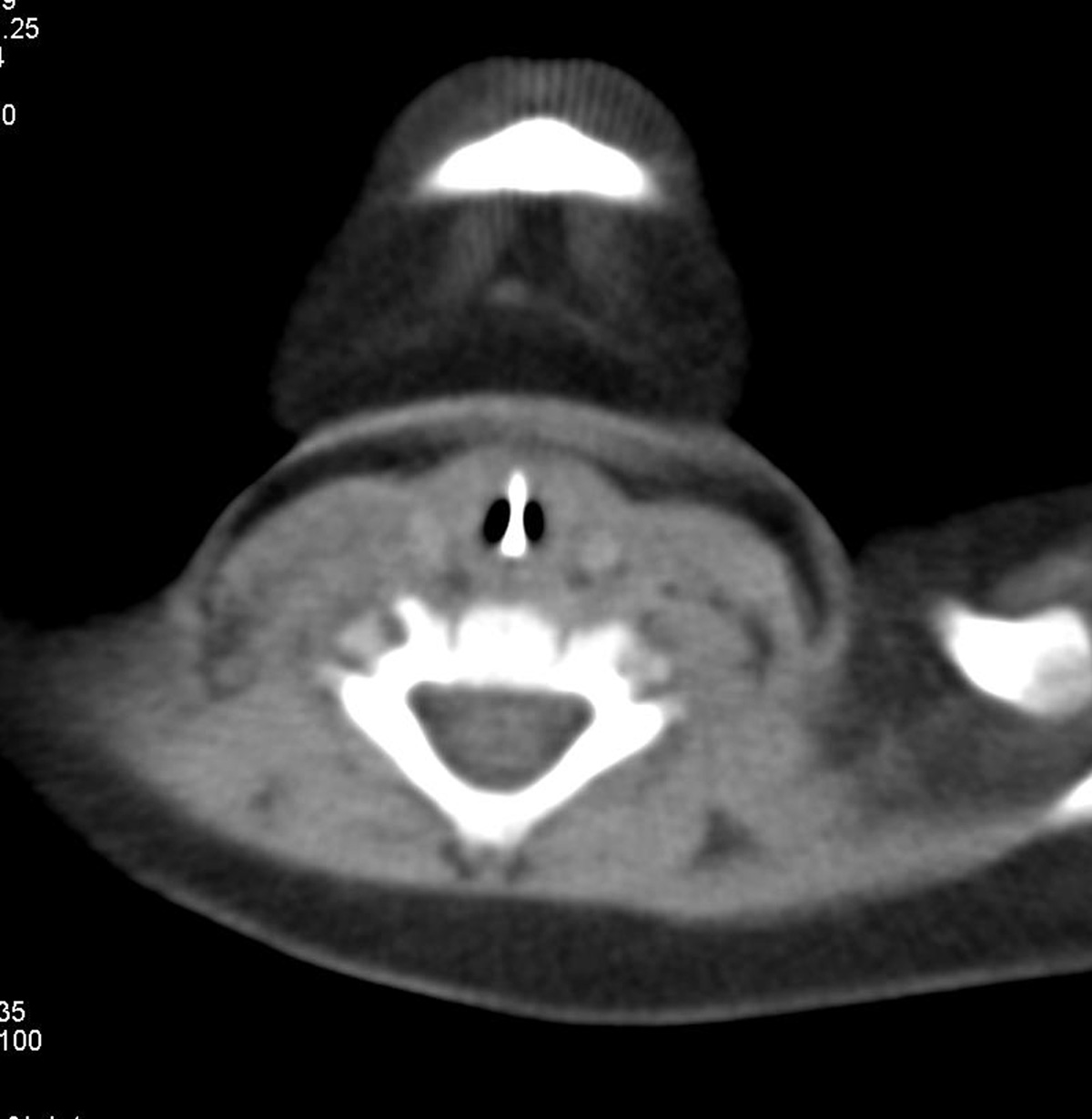
Tracheal Foreign Body - CT Scan with 3D.

CT scan axial soft tissue window of 7 Months old male child with history of foreign body aspiration with asymptomatic baby showing linier metallic foreign body oriented vertically in the center with patent lumen on either side. 

Coronal reformatted CT scan soft tissue window well delineate the vertical extent of the foreign body

Minimum intensity projection (minIP) image showing the foreign body. 

3D Volume rendered image showing showing the foreign body. 

Virtual bronchoscopy showing showing the foreign body (arrow). 

Extraction by bronchoscopy is the treatment of choice for tracheal foreign bodies

Friday, July 1, 2011

Healed Varicella Pneumonia


There are multiple tiny subcentimeter calcific densities noted throughout both lung parenchyma more on the left side. There is no focal lung parenchymal mass or cavitating lesion seen. The appearances are classical for previous varicella exposure. 

Reference: 
Kim EA, et al. Viral Pneumonias in Adults: Radiologic and Pathologic Findings. RadioGraphics 2002; 22: 137.Credit: Dr Abhijit Datir

Monday, May 2, 2011

Carcinoma lung with intra-spinal metastases - PET/CT.

PET/CT fused image of a 65 year old smoker showing large intensely FDG avid mass lesion in the left upper lobe with areas of necrosis and mediastinal FDG avid lymphadenopathy.

Sagittal PET/CT fused whole spine image showing intraspinal intense FDG avid focal lesions at C7-D1 and L1 levels suggestive of metastases.

Sagittal PET/CT fused cervical spine image showing intraspinal intense FDG avid focal lesion.

Axial PET/CT fused cervical spine image at C7 level showing intraspinal intense FDG avid focal lesion.

Axial PET/CT fused cervical spine image at L1 level showing intraspinal intense FDG avid focal lesion

Discussion:

The intraspinal metastases may spread through several routes, such as arterial dissemination; venous spread through the network of communicating venous channels in the spinal cord and the cauda equina; spread via the perineural lymphatic vessels or the subarachnoid space; invasion of a dorsal root; or direct extension of the primary tumor.
Lung carcinoma commonly metastasize by arterial dissemination.

Wednesday, September 29, 2010

The Azygos Lobe

55 year old male came with history of chronic cough and was reffered for CT scan to ruleout tuberculosis and any other lung disease. CT scan axial section (first image) shows a azygos vein is coursing through the apical segment of the right upper lobe (arrow). Coronal reformated image shows pleural reflection - mesoazygos (arrow) with azygos vein (arrow head) in the right apical region.

Discussion:

The azygos lobe is a rare anomaly that developmentally arises when the right posterior cardinal vein penetrates the apex of the lung, instead of passing over it, and travels inferiorly taking pleural layers with it to entrap a portion of the right upper lobe. The two folds of pleura form the mesoazygos, a fissure visible on 0.4% of chest radiographs and 1.2% of high resolution computed tomography (CT) studies. The right azygos lobe is supplied by the medial segments of the apical and anterior or posterior branches of the apical segmental bronchial artery and vein. A true left azygos lobe has also been reported.
On chest radiography, the azygos lobe is usually distinguished by the azygos fissure, which superiorly has a triangular shape and inferiorly demonstrates the azygos vein as a tear-shaped shadow. The azygos fissure typically appears as a fine, convex line that crosses the apex of the right lung. The azygos lobe can appear opaque and be incorrectly interpreted as a pathologic mediastinal finding on PA chest radiographs.
When findings on traditional imaging are not clear, CT exams can be helpful in delineating relevant anatomy. Clinically, the knowledge of azygos lobe anatomy is important during thoracic surgical approaches. Partial obstruction of the thoracoscopic view during a bilateral sympathectomy was reported during attempted mobilization of the azygos lobe. Others reported difficulty reflecting the pleura during primary repair of the esophageal atresia in a pediatric patients. There are also reports of the phrenic nerve coursing within the azygos fissure. Finally, multiple authors have reported spontaneous pneumothorax associated with the azygos lobe in both the adult and the pediatric patient.

Sunday, January 24, 2010

Disseminated tuberculosis - CT


Disseminated tuberculosis (TB) is a contagious bacterial infection that has spread from the lungs to other parts of the body through the blood or lymph system. The differential diagnosis of miliary nodules in bilateral lungs are tuberculosis, metastases, sarcoidosis, lymphoma, hypersensitivity pneumonitis and rare causes like silicosis, beryliosis, stanosis. Depending imaging findings and patients clinical deatils, one can differentiate each other.
The differential diagnosis of patients with fever, splenomegaly, and multiple hypoechoic hypoattenuating splenic lesions includes splenic abscesses that may be bacterial, fungal, or granulomatous in nature. Pyogenic abscesses are usually solitary. On US images, they appear as small irregular hypoechoic lesions that may evolve into a poorly defined heterogeneous mass. On CT images, they typically appear as single irregularly marginated hypoattenuating lesions. The presence of intralesional gas is pathognomonic of pyogenic infection, although it occurs infrequently. Multiple splenic abscesses, usually smaller than 2 cm in diameter, are commonly associated with nonbacterial infections (ie, fungal and granulomatous infections). Fungal abscesses, when visible, characteristically appear on US images as round hypoechoic lesions with a central hyperechoic area (ie, “bull's-eye” or “target” appearance) that corresponds to inflammatory cells with a surrounding hypoechoic band of fibrosis. The “wheel-in-a-wheel” pattern is seen when the central portion becomes necrotic and hypoechoic. Contrast-enhanced CT typically depicts fungal splenic abscesses as multiple small low-attenuation lesions. Patients with cat-scratch disease may also develop splenomegaly and splenic microabscesses. Neoplasms that involve the spleen—such as lymphoma (both Hodgkin and non-Hodgkin lymphoma), leukemia, metastasis, or even Langerhans cell histiocytosis—may manifest as focal hypoechoic hypoattenuating lesions. Sarcoidosis, which is a systemic granulomatous disease of unknown origin, may also have abdominal involvement with lymphadenopathy and hypoattenuating nodules in the liver and spleen. It is generally impossible to differentiate visceral TB lesions from fungal infections, lymphoma, or metastasis—unless they are associated with other typical abdominal TB findings, such as lymph node or bowel involvement.

Saturday, January 9, 2010

Bronchocele - CT

Chest scanogram of 23 year old male patient came with history of cough showing eliptical ares of increased opacity seen in the right upper lobe, medial end pointing towards the hilum and the lesion is sharply marginated.
Contrast enhanced CT scan showing non enhancing fluid attenuating lesion seen in the right upper lobe appears to be with in the dilated bronchi of posterior segment of right upper lobe. Can better appreciate on coronal reformatted image.

Bronchocele is commonly seen in congenital bronchial atresia, where there will be fluid accumulation in the bronchus distal to the atretic segment of bronchus. There are other rare causes for bronchocele like tumor obstructing the segmental bronchus, commonly carcinoids. The distal involved alveoli may show hypoplasia or a probable consequence of decreased ventilation and perfusion. These alveoli however remain open by collateral air drift occurring by the interalveolar pores of Kohn (air drift) or the bronchoalveolar channels of Lambert or the interbronchiolar channels with some amount of air trapping. Although radiograph is the first investigation to be done and suspected on it. The findings may show as described above in our case. Spiral CT is the examination of choice, not only to show all the components of the anomaly and to estimate the extent of air trapping but also for ruling out differential diagnosis such as bronchogenic cyst, bronchiectasis, aspergillosis, completely thrombosed arteriovenous malformation or pulmonary aneurysms and tumors.

Saturday, November 28, 2009

MEDIASTINAL NEUROBLASTOMA

One year old female baby came with history of persistent cough, difficulty in breathing and stridor. Baby also had weight loss and sometimes mild fever. Routine hematological investigations were normal. Chest radiograph and contrast enhanced CT scan showed below mentioned findings

Figure 1 a) Radiograph frontal view showing homogenous opacity lesion in the right upper and mid zone with broad base towards mediastinum. The lesion is causing shift of mediastinum and trachea to the contralateral side and compressing the trachea just above the carina (arrow head). The lesion is extending above the clavicle (cervico-thoracic sign) and erosion of posterior end of third and fourth ribs (Long arrow). C) Contrast enhanced CT scan axial section mediastinal window showing heterogeneously enhancing mass lesion in the right posterior mediastinum extending to middle mediastinum causing compression of the trachea. The lesion is showing multiple small foci of calcification (black arrows in fig c).

Discussion:

Primary mediastinal neuroblastoma accounts for 14% of all neuroblastomas. Common location is posterior mediastinum, where they arise from paravertebral sympathetic chain. Patient commonly present with chest pain, cough and respiratory distress due to airway obstruction. Chest radiograph is the initial radiological investigation to be done in such patients. It demonstrates homogenous opacity mass in the mediastinum. If the tumor is large, there may be extrinsic compression and displacement of trachea and main bronchus with mediastinal shift (fig a-c). Rib erosion and asymmetrical widening of inercostal spaces may be seen on radiograph. Intraspinal extension may be seen as loss of vertebral height with widening of neural foramina on lateral radiograph. Approximately 50% of the lesions show calcification on radiograph.

CT scan is usually done to confirm the presence of lesion, and it also helps to define the location and extent of lesion, adjacent organ involvement, or vascular involvement. CT demonstrates calcification in up to 90%. The lesion characterization has become better with the advent of multiplanar image reconstructions in MDCT. However MR imaging is the investigation of choice for better characterization and demonstration of full extent of mass, extradural intraspinal extension and chest wall invasion. The advantages of MR over CT are its direct multiplanar imaging and better contrast resolution, which can allow differentiation of vascular from non vascular lesions without the aid of intravenous contrast agent. MR is very often used to evaluate tumor involving spine and spinal canal.

Teaching point:

Radiograph is the initial investigation to be done in the mediastinal masses and one should be able to identify the lesion and arrive at differential diagnosis. CT scan and MRI are done to confirm the lesion, location, extent, adjacent organ involvement or vascular involvement and intraspinal extension.

Saturday, August 8, 2009

Pulmonary alveolar proteinosis (PAP)

HRCT of 30 year old female shows welldefined ground glass opacities which is associated with marked intelobular septal thickening and a sharp nonanatomic demarcation between normal and abnormal lung suggestive of "Crazy paving" pattern which is typical of PAP.

Pulmonary alveolar proteinosis (PAP) is a rare (1 case per 2 million), diffuse lung disease that is characterized by the alveolar and interstitial accumulation of a periodic acid-Schiff (PAS) stain-positive phospholipoprotein that is derived from surfactant.The lung architecture is otherwise normal, and any associated inflammation or fibrosis is limited in extent. Pulmonary alveolar proteinosis (PAP) occurs in primary and secondary forms. Primary PAP is either idiopathic (90% of cases) or congenital (2% of cases) in origin, whereas secondary PAP occurs in association with various pathologies like hematologic malignancies, particularly chronic myeloid leukemia and lymphomas, occupational exposures and associated with infections. Congenital PAP results from deficiency of surfactant protein B (SP-B) or abnormality of the granulocyte-macrophage colony-stimulating factor (GM-CSF) receptor β chain.
CT scan with the above findings are almost diagnostic. razy paving is the characteristic finding of PAP on high-resolution CT (HRCT) scanning and consists of patchy, bilateral, geographic areas of ground-glass opacity that are associated with interlobular septal thickening. The disease is often distributed uniformly from the lung apex to the base. Interlobular septal thickening may be encountered more frequently in the lower lung zones.Classically, the abnormal pulmonary parenchyma is demarcated sharply from normal lung areas without a discernible anatomic boundary.
The crazy paving pattern also seen in many other condition like hydrostatic pulmonary edema, diffuse alveolar damage from any number of causes, pulmonary hemorrhage, diffuse pulmonary infections (including M tuberculosis, M pneumoniae, and other bacterial pneumonias), the diffuse form of bronchioloalveolar carcinoma, ARDS, drug-induced pneumonitis, radiation pneumonitis, bronchiolitis obliterans organizing pneumonia, chronic eosinophilic pneumonia, obstructive pneumonitis, acute interstitial pneumonia, and lipoid pneumonia.

Search This Blog