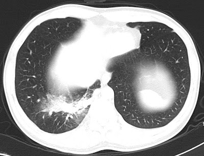
31 year old male patient came with repeated respiratory tract infections. CT showed consolidation in right lower lobe. Mediastinal window images show suspicious small artery supplying the lesion from the descending thoracic aorta for which CT angio was done
CT angio MIP and VRT images showing large single artery supplying the sequestrated segment of the lung. Venous drainage to the pulmonary veins. Later patient was taken for angioembolization of aberrant artery.
Discussion:
Definition: An aberrant lung tissue mass that has no normal connection with the bronchial tree or with the pulmonary arteries.
Arterial blood supply: Systemic arteries, usually the thoracic or abdominal aorta.
Venous drainage: Azygous system, the pulmonary veins, or the inferior vena cava.
Two types:
1. Intralobar sequestrations: can manifest as an area of increased opacity simulating pneumonia, as a mass with or without air-fluid levels, or as cysts.
2. Extralobar sequestration, in which the sequestered lung has its own separate pleural covering, is much less common and usually is found on the left side next to the hemidiaphragm. At radiography, it may manifest as a reasonably well-defined mass at the base of the left hemithorax. Rarely, an intralobar and extralobar sequestration may occur in the same patient.
The main differentiating points are as given in the following table.


1 comment:
Nice post
book CT scan in Delhi with the high quality diagnostic labs via easybookmylab
Post a Comment