
HRCT of right temporal bone axial image showing transverse temporal bone fracture (arrow) and the fracture line passing through the first anterior genu of the facial nerve canal (arrow head).
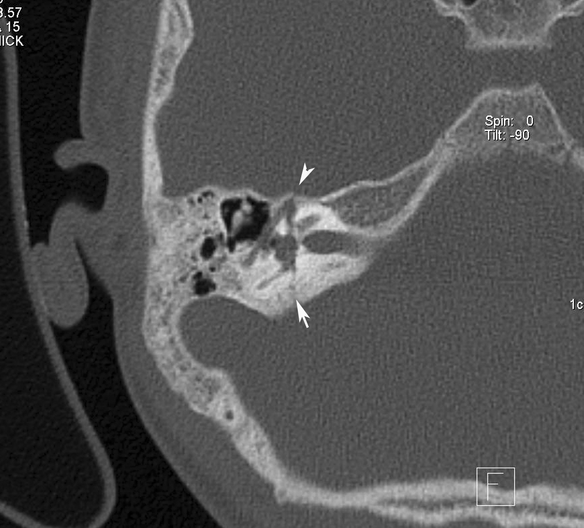
Subsequent section of the HRCT right temporal bone showing the fracture passing through the facial nerve canal (arrow head).
Classification:
1. Longitudinal (parallel to the axis).
2. Transverse (perpendicular to the axis).
3. Oblique/Mixed- Common.
Longitudinal fracture: The fracture line of force runs roughly from lateral to medial. A fracture line may extend through the facial nerve canal, thereby damaging the facial nerve. Associated injury, such as transection or intraneural hemorrhage, may cause facial nerve paralysis, as can damage from displaced bone fragments.
Mixed fractures: Oblique (or mixed) fracture patterns, which extend both longitudinally and transversely, are common, and some case series report that these occur more often than do isolated transverse or longitudinal fractures.
Radiology:
Radiography: Plain film radiographs of the skull may show opacified mastoid air cells, intracranial air, or, rarely, a lucency (fracture line).
HRCT: can demonstrate a lucency through the temporal bone. Involvement of the middle ear, petrous bone, otic capsule, and facial nerve canal are the primary determinants of prognosis.
MRI: may demonstrate fluid (high signal on T2-weighted images) in the middle ear and mastoid air cells. T1-weighted images may reveal a bright signal in the labyrinth or middle ear, consistent with hemorrhage.

No comments:
Post a Comment