Friday, October 28, 2011

Carcinoma pancreas - USG and CT

60 Year old female with history of pain abdomen and weight loss. USG showing well defined hypoechoic mass lesion in the region of body and tail of the pancreas with mild to moderate vascularity. It also shows few necrotic areas.

Axial CT scan showing well defined heterogeneously enhancing mass lesion arising from the body and tail of the pancreas showing areas of necrosis and causing mass effect on the adjacent structures however no obvious infiltration of the surrounding structures.

USG Guided FNAC showing the lesion with echogenic linier needle (arrow head)

Discussion:

Pancreatic adenocarcinoma is the second most common cause of death from GI cancers. 
Adenocarcinoma ususally arise from pancreatic ducts (99%) and other site being acinar cells (1%).
More than 90% of pancreatic cancers appear in the late stage of disease.

Role of radiology:
1. Early detection.
2. Determination of resectability of the tumor.
3. Relationship to surrounding vasculature.

Multiphasic contrast enhanced study of upper abdomen in Multislice CT scan is the preferred investigation of choice.
CT Scan features include:
1. Alterations in morphology of the gland.
2. Obliteration of peripancreatic fat.
3. Loss of sharp margins with surrounding structures.
4. Involvement of adjacent vessels - is the sign of unresectability.
5. Regional lymph node enlargement.
5. Pancreatic ductal dilatation, pancreatic atrophy, and obstruction of the common bile duct (CBD)

MRI: Role of MRI is yet to be firmly established. T1-weighted fat-suppressed spin-echo and single–breath-hold gradient-echo fast low-angle shot (FLASH) sequences with gadolinium enhancement are valuable for tumor detection.

USG:
The lesion may have a variable appearance on US. It may be hypoechoic, isoechoic, or hyperechoic to the normal pancreas.
Pancreatic ductal dilatation and biliary ductal dilatation are easily demonstrated in patients with a tumor in the head of pancreas that causes an obstruction.
Lymphadenopathy, the relation of the tumor to peripancreatic vessels, and the tumor margins are demonstrated less reliably with US than with other modalities.

Monday, October 17, 2011

Unusual migration of proximal tip of ventriculoperitoneal shunt - CT

26 year old male patient with post traumatic hydrocephalus treated with ventriculoperitoneal shunt axial CT sectio at hte level of third ventricle showing tubular hyperdense structure (arrow) suggestive of VP shunt.

Axial section at the level of basal cisterns showing the VP shunt catheter in the suprasellar structures.

Sagittal reformatted image showing the catheter tip is situated at the roof of the sella.

Discussion:
  • There are many incidents of unusual migration of the shunt tip in to various parts of the body. 
  • Usually it is the distal or the peritoneal catheter that migrates after breakage or disconnection into many locations such as the scalp, heart, anus/rectum, urethral, knee, umbilicus, chest, pleural cavity, inguinal canal and scrotum.
  • Complete migration of the shunt in to the ventricles have been reported.
  • This might be the first case of shunt migration in to the suprasellar region as far as my knowledge is concerned.

Sunday, October 9, 2011

Pottt's spine with bilateral psoas abscess - MRI


Sagittal T2 and T1 images of MRI lumbar spine in 20 year old male showing partial collapse of the L4 vertebral body with restropulsion of the posterior fragment causing thecal sac compression and mild compression on the traversing nerve roots. 

Axial T2 image at the level of L4 showing bilateral large psoas abscesses.
Coronal image well depict the craniocaudal extent of the abscess.

Lipoma Arborescens of the Knee - MRI

Sagittal T1 weighted MRI in 27 year old male shows a large suprapatellar effusion with a frond-like synovial mass of fat intensity in suprapatellar region and in posterior intercondylar region (arrows).
Sagittal PD FS MRI shows the synovial mass to be the same low intensity as fat (arrows).

Axial gradient echo MRI shows the characteristic frond-like pattern.


Discussion:
Lipoma arborescens is a rare benign lesion, which arises in the synovium and is characterized by villous proliferation of fat cells. The proliferating cells appear organic and often resemble a tree thus its name, arborescens, from the Latin arbor for tree.

Cause: is uncertain. One hypothesis is - synovial hyper-proliferation is in response to traumatic or inflammatory stimuli.
Associations: Trauma, osteoarthritis or rheumatoid arthritis.

Radiograph: Findings are non specific.
1. Soft tissue density with areas of lucency suggesting fat.
2. Underlying degenerative changes of the knee.

MRI: is the investigation of choice.
  • Villous proliferation, often with a frond-like configuration, which arises from within the synovial cavity.
  • Fatty characteristics and typical pattern of proliferation shows isointense to the fat on all the sequences.
  • Joint effusion.
  • Meniscal  tears.
  • Baker's cyst in 38%.

Intralobar sequestration - CT Angiography.

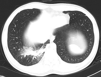
31 year old male patient came with repeated respiratory tract infections. CT showed consolidation in right lower lobe. Mediastinal window images show suspicious small artery supplying the lesion from the descending thoracic aorta for which CT angio was done


CT angio MIP and VRT images showing large single artery supplying the sequestrated segment of the lung. Venous drainage to the pulmonary veins. Later patient was taken for angioembolization of aberrant artery.

Discussion:

Definition: An aberrant lung tissue mass that has no normal connection with the bronchial tree or with the pulmonary arteries. 
Arterial blood supply: Systemic arteries, usually the thoracic or abdominal aorta.
Venous drainage: Azygous system, the pulmonary veins, or the inferior vena cava.

Two types:  
1. Intralobar sequestrations: can manifest as an area of increased opacity simulating pneumonia, as a mass with or without air-fluid levels, or as cysts. 
2. Extralobar sequestration, in which the sequestered lung has its own separate pleural covering, is much less common and usually is found on the left side next to the hemidiaphragm. At radiography, it may manifest as a reasonably well-defined mass at the base of the left hemithorax. Rarely, an intralobar and extralobar sequestration may occur in the same patient.
The main differentiating points are as given in the following table.


Thursday, October 6, 2011

Scapholunate ligament tear - MRI

Radiograph of wrist in 45 Year old male patient with pain in the wrist joint showing widened scapholunate joint space with focal periosteal elevation in the lunate (arrow).

Coronal PD FS image showing complete tear in the scapholunate ligament with mild marrow edema in the scaphoid and lunate bones.

Coronal GRE image well depicts the ligament tear (arrow).
Discussion:

  • Most common and most significant ligament injury of wrist causing carpal instability.
  • Risk factors: ulna minus configuation, slope of radial articular surface, and lunotriquetral coalition.
Spectrum of injury include: (increasing severity)
1. Dynamic scapholunate instability
  • No radiographic evidence of malalignment is present (ie dynamic deformity); 
  • Diagnosis is established by dorsal S-L tenderness and positive shift test; 
2. Rotatory subluxation of scaphoid:
3. Scapholunate dissociation (SLD):

  • SL ligament tear may lead to rotational dislocation of scaphoid allowing proximal pole to displace posteriorly & distal pole to displace anteriorly.
  • Scaphoid inherently tends to palmar flex because of its oblique position and the loading applied through (STT) joint.
  • Because scaphoid lacks proximal of ligament, it will rotate around radiocaptitate ligament leading to dorsal rotary subluxation of the proximal pole.
4. Dorsal intercalated segment instability: (DISI) 
5. Scapholunate advanced collapse:

Osteosarcoma of femur - Radiograph and MRI

Radiograph of 20 year old female showing destructive lytic lesion involving the metaphysis of the medial aspect of femur with new bone formation and periosteal reaction (caudman triangle) {Arrow}

T1 weighted coronal image showing lytic destructive lesion with soft tissue, periosteal reaction and new bone formation.

Axial GRE image showing the new bone formation as hypointense signal and periosteal reaction as hyperintense signals.

Coronal STIR image showing the lesion.
Discussion: Osteosacroma is malignant tumor of bone in which neoplastic osteoid is produced by a proliferating spindle cell stroma.

  • Most common primary malignant bone tumor of mesenchymal derivation.
  • Arises in adolescents, and second or third decade.
  • Affects males slightly more often than females. 

Affected sites:
  • Occurs in region of knee (distal femur or proximal tibia) in 50% of patients. 
  • Other sites include proximal humerus, proximal femur, & pelvis.
  • Most osteosarcomas occur in the metaphysis.
  • Infrequently occurs in the spine.
Classification:
  1. Classic 
  2. Telangiectatic 
  3. Parosteal 
  4. Periosteal
IMAGING:
Radiograph:
  • Destructive lytic lesion or mixture of lytic and sclerotic areas (common).
  • Moth eaten appearance with ill-defined zone of demarcation.
  • Involves the metaphysis
  • New bone formation.
  • Periosteal reaction
  • Visible soft-tissue mass.
  • Spiculated / Sunburst appearance.
  • 'Codman's Triangle' which is basically a subperiosteal lesion formed when the periosteum is raised due to the tumor.
CT Scan:
  • Gives clearer indication of bone destruction.
  • May depict small amounts of mineralized osseous matrix not seen on radiographs. 
  • May be particularly helpful in visualizing flat bones, in which periosteal changes may be more difficult to appreciate.
MRI:
  • Modality of choice in evaluating the local extent of disease because of its excellent bone marrow and soft tissue contrast and multiplanar capabilities.
  • it assists in determining the most appropriate surgical management.
  • Better delineates the involvement of the epiphysis, adjacent joint, adjacent soft tissues, neurovascular bundle, marrow extent and skip lesions.
Bone Scans:
  • Increased uptake of radioisotope on bone scans obtained by use of technetium-99m (99m Tc) methylene diphosphonate (MDP).
  • most useful in excluding multifocal disease but skip lesions are more reliable on MRI.

Wednesday, October 5, 2011

Giant Cell Tumor of Fibular head - Radiograph and MRI.

Radiograph of the knee in 32 year old male patient showing well defined expansile lytic lesion seen in the  epimetaphyseal region of fibular head with thinning of the cortex and cortical break at few places.

Axial T2 FS image at the level of fibular head showing expansile lytic lesion showing fluid contents with septations, thinning of the cortex and cortical break at few places. No obvious blood fluid levels noted.

Axial T1 weighted image at the level of fibular head showing the lesion with isointense contents and thinning of the cortex.

Coronal T1 weighted images showing the lesion.
Discussion:
  • GCT is a common benign but locally aggressive lesion of unknown etiology. 
  • It occurs chiefly in men between 20-50 yrs (after epiphyseal closure).
  • The tumor is expansile lytic lesion that involves the epiphysis & metaphysis.
  • The tumor may enlarge to occupy most of epiphysis & adjacent metaphysis. 
  • The tumor may erode & penetrate subchondral bone, articular cartilage, & cruciate ligaments.
Location:

  • Epiphysis of distal femur, proximal tibia, & distal radius.
  • Other sites: fibula, sacrum, proximal humerus, & distal tibia.
  • Can occur in bones of pelvis, particularly ilium near SI joint and sacrum.
Spine:
  • Usually located in vertebral body; 
  • Radiolucent lesion in vertebral body of a young patient is likely to be GCT; 
Staging:
Stage I:
  • Benign latent giant cell tumors.
  • No local aggressive activity.
Stage II:
  • Benign active GCT. 
  • Imaging studies demonstrate alteration of the cortical bone structure.
Stage III:
  • Locally aggressive tumors.
  • Imaging studies demonstrate a lytic lesion surrounding medullary and cortical bone.
  • There may be indication of tumor penetration through the cortex into the soft tissues.
IMAGING:
Radiographic Features: well-defined lytic lesion that involves the metaphysis and epiphysis (typical of a giant cell tumor);

CT Scan: helps to determine the extact amount of cortical destruction and helps determine the optimal location of the cortical window;

Bone Scans: Bone scans may show decreased radioisotope uptake in the center of lesion (doughnut sign). It is also found in ABC.

MRI:
  • MRI May show fluid fluid or blood fluid levels.
  • Help determine determine extent of tumor destruction and soft tissue involvement.
  • May be indicated when the tumor has eroded through the cortex and allows determination of whether concomitant neurovascular structures are involved.
  • May help evaluate subchondral penetration.
Differential diagnosis:
1. Aneurysmal bone cyst - ABC.
2. Non Ossifying fibroma.

Search This Blog