Friday, April 1, 2011
Frontoethmoidal Encephalocele
Wednesday, February 16, 2011
Tibialis posterior tenosynovitis - Ultrasonography
48 year old male came with history of redness and swelling in the posteromedial aspect of the right ankle. Ultrasonography showed mildly thickened tibialis posterior tendon with significant amount of fluid collection around the tendon. No effusion in the ankle joint/ No DVT / No varicose veins. Features are consistent with tibialis posterior tenosynovitis.
Discussion:
Tenosynovitis is inflammation of tendon sheath. It can be caused by trauma, pyogenic infection, tuberculosis, seronegative spondylarthropathies or rheumatoid arthritis.
- Thickened tendon.
- Fluid collection around the tendon.
- Mildly increased vascularity within the tendon.
- Fluid collection around the tendon in its proximal aspect.
- Signal change within the tendon is usually not seen in the acute stage and will be seen in chronic stage.
Monday, January 31, 2011
Neurocysticercosis - MRI
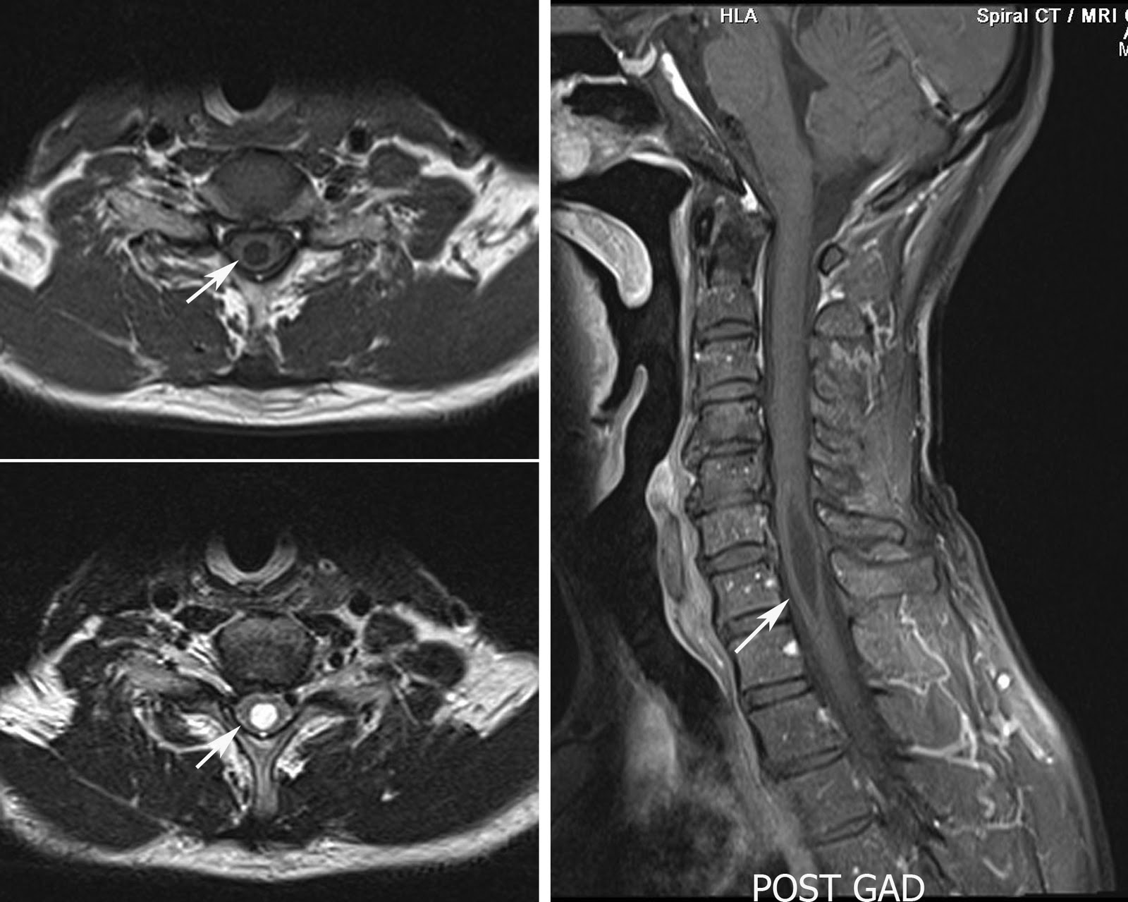
Syrinx - MRI
- Communicating (with the subarachnoid space, usually at the level of the obex at the inferior aspect of the fourth ventricle)
- Posttraumatic
- Tumor-related
- Arachnoiditis-related
- Idiopathic.
- Chiari II malformation with hydrocephalus
- Chiari I malformation without hydrocephalus
- Extramedullary compressive lesions at the craniocervical junction or along the length of the spinal canal
- Spinal cord trauma
- Intramedullary tumors and intraperimedullary infections
- Multiple sclerosis
Tuesday, January 25, 2011
Endometrioma - MRI
- A round-shaped homogeneous hypoechoic 'tissue' of low-level echoes without papillary proliferations associated with 'poor' vascularization (defined by Aleem et al. in 1995);
- A round-shaped homogeneous hypoechoic 'tissue' of low-level echoes with an echogenic portion in which no flow is detected (atypical findings).
Thursday, January 13, 2011
HIV Encephalopathy - MRI
Choledocholithiasis - MRCP
USG: Sensitivity varies from 13 to 55%. Features include dilated duct with stones visualized within. Recently endoscopic ultrasonography (EUS) has also been used with very high sensitivity and specificity.
CT: Moderate sensitivity of 65 - 88% but have to look for very subtle findings. They are
- Target sign - Central rounded density of stone with surrounding lower attenuating bile or mucosa.
- Rim sign: Stone is outlined by thin shell of density.
- Crescent sign: Bile eccentrically outlines luminal stone, creating a low attenuation crescent.
- Calcification of the stone: unfortunately only 20% of stones are of high density
MRCP: is the gold standard investigations for biliary lithiasis replacing the ERCP. Filling defects are seen within the biliary tree on thin cross-sectional T2 weighted imaging.
Tuesday, January 11, 2011
Carcinoma Prostate - PET/CT
- Fluorodeoxyglucose (FDG): have a role in the detection of lymph node metastases, particularly in patients with relapsed disease after primary treatment. The sensitivity is around 50%. FDG-PET has an excellent detection rate for lytic skeletal metastases, but it has a poor detection rate for sclerotic metastases as in our case.
- Carbon 11 (C11)–acetate and C11-choline (18F-Choline): Shown promising alternative but they are less readily available and still under assessment. Few recent studies have showed conflicting results as one study says there is relative high rate of false-negative results as prostatic disorders other than cancer may accumulate 11C-choline. Some other study shows C11-choline PET/CT can differential prostate carcinoma from benign hyperplasia, chronic prostatitis, or normal prostate tissue. The C11 choline has shows promising results in staging of the tumor; however it still needs more studies.
Thursday, December 30, 2010
Cervical Carcinoma-MRI
- Can quantify the volume of the tumor especially in the early stages.
- Metastatic lymphnode evaluation.
- Obviates the use of invasive procedures like cystoscopy and proctoscopy.
- Brachytherapy and external beam radiation therapy can be optimised with the MR imaging.
- Detect and confirm invasion of the adjacent organs.
- Identify fistulous tracts if any.
Thursday, December 16, 2010
Bone infarct - MRI
- Stage 0 - Normal findings are demonstrated.
- Stage I - The appearance may vary from normal to subtle trabecular mottling, but an isotopic bone scan or MRI shows abnormal bone.
- Stage II
- Stage IIa - Focal radiopacity is associated with osteopenia.
- Stage IIb - Radiopacity is associated with osteoporosis and an early crescent sign.
- Stage III
- Stage IIIa - An established crescent sign is associated with cyst formation.
- Stage IIIb - Mild alteration in the configuration of the femoral head is caused by a subchondral fracture, but the joint space is maintained.
- Stage IV - Marked collapse of the femoral head is demonstrated with an associated acetabular abnormality.
- Stage V - Joint space narrowing is demonstrated with changes of secondary osteoarthrosis.
Saturday, December 11, 2010
Achilles Tendonitis - MRI


46 Year old male with pain in the left ankle MRI T2 FS sagittal and T2 FSE axial images showing diffuse hyperintensities in the distal Achilles tendon with mild marrow edema in the calcaneous at the insertion site and mild peritendinous inflammatory changes suggestive of Achilles Tendinitis.
Thursday, September 30, 2010
Cholesteatoma- HRCT
Non contrasted temporal bone CT reveals a soft tissue mass in the right middle ear and Prussak’s space with associated erosions of the scutum, epitympanum walls, lateral semicircular canal, tegmen tympani and middle ear ossicles. It is also extending in to the mastoid air cells.- An acquired cholesteatoma is a collection of exfoliated squamous epithelium and debris.
- Cholesteatomas often begin at the pars flaccida of the tympanic membrane and grow in Prussak's space of the temporal bone and produce inflammatory reaction.
- Cholesteatomas typically occur in the setting of chronic dysfunction of the Eustachian tubes and recurrent otitis media.
- Cholesteatomas can be as small as a couple of millimeters in size or can grow to fill the entire middle ear.
- Erosions of the scutum and ossicles are commonly seen.
- The presence of bony erosions or expansion are strong support of cholesteatoma over chronic otomastoiditis.
- Acquired cholesteatomas occur in children and adults.
- Most common presenting symptoms include ear discharge, hearing loss, ear pain and vertigo.
- Without treatment cholesteatomas will progressively increase in size.
- Complications include: Hearing loss, CN 7 palsy, venous sinus thrombosis, semicircular canal fistulas, and intracranial invasion.
- Early surgical intervention usually results in complete eradication and preservation of hearing.
- Acquired cholesteatoma
- Chronic otomastoiditis
- Middle ear cholesterol granuloma
- Glomus tympanicum paraganglioma
Wednesday, September 29, 2010
Double left renal artery - CT Angiography
44 Year old female referred for CT renal angiography for transplant donor evaluation. The coronal volume rendering images showing normal bilateral kidneys with single right renal artery and two renal arteries on the left side.Discussion:
CT angiography is the most preferred investigation for donor kidney evaluation since it gives excellent anotomical depiction of the vessels, organs and the function of kidney in urography.
Protocol - Arterial phase, Venous phase and Excretory phase.
The things to be mentioned in the radiological report are
- Size, shape and position of the kidneys.
- Number of renal arteries, any accesory artery.
- Course of left renal vein retroaortic /preaortic.
- Distance of renal artery from origin to first branching.
- Length of right renal vein.
- Any other normal varuients/renal masses.
- Collecting system - Duplication of any.
All these findings are valuable to the graft harvesting surgeon and for the transplant anastomosis.
The Azygos Lobe
55 year old male came with history of chronic cough and was reffered for CT scan to ruleout tuberculosis and any other lung disease. CT scan axial section (first image) shows a azygos vein is coursing through the apical segment of the right upper lobe (arrow). Coronal reformated image shows pleural reflection - mesoazygos (arrow) with azygos vein (arrow head) in the right apical region.Discussion:
The azygos lobe is a rare anomaly that developmentally arises when the right posterior cardinal vein penetrates the apex of the lung, instead of passing over it, and travels inferiorly taking pleural layers with it to entrap a portion of the right upper lobe. The two folds of pleura form the mesoazygos, a fissure visible on 0.4% of chest radiographs and 1.2% of high resolution computed tomography (CT) studies. The right azygos lobe is supplied by the medial segments of the apical and anterior or posterior branches of the apical segmental bronchial artery and vein. A true left azygos lobe has also been reported.
On chest radiography, the azygos lobe is usually distinguished by the azygos fissure, which superiorly has a triangular shape and inferiorly demonstrates the azygos vein as a tear-shaped shadow. The azygos fissure typically appears as a fine, convex line that crosses the apex of the right lung. The azygos lobe can appear opaque and be incorrectly interpreted as a pathologic mediastinal finding on PA chest radiographs.
When findings on traditional imaging are not clear, CT exams can be helpful in delineating relevant anatomy. Clinically, the knowledge of azygos lobe anatomy is important during thoracic surgical approaches. Partial obstruction of the thoracoscopic view during a bilateral sympathectomy was reported during attempted mobilization of the azygos lobe. Others reported difficulty reflecting the pleura during primary repair of the esophageal atresia in a pediatric patients. There are also reports of the phrenic nerve coursing within the azygos fissure. Finally, multiple authors have reported spontaneous pneumothorax associated with the azygos lobe in both the adult and the pediatric patient.
Tuesday, August 3, 2010
Adrenal Myelolipoma - CT
CT scan of a 25 year old female showing well defined fat containing lesions seen in the region of right adrenal gland (arrow) suggestive of adrenal myelolipoma.