Saturday, March 16, 2013

Lung Abscess


45 year old male with history of high grade fever and foul smelling expectorations. Chest radiograph showed well defined thick walled cavitory lesion in the right lower lobe with air fluid levels consistent with lung abscess.

Discussion: 

A lung abscess is a circumscribed collection of pus within the lung, is are potentially life threatening. They are often complicated to manage and difficult to treat.


A primary abscess is one which develops as a result of primary infection of the lung. They most commonly arise from aspiration, necrotising pneumonia or chronic pneumonia, e.g. pulmonary TB


A secondary abscess is one which develops as a result of another condition. e.g: bronchial obstruction: bronchogenic carcinoma, inhaled foreign body.


Plain Radiograph: The classical appearance is a cavity containing an air-fluid level. In general abscesses are round in shape, and appear similar in both frontal and lateral projections. Additionally all margins are equally well seen, although adjacent consolidation may make assessment of this difficult.

CT scan:

CT is the most sensitive and specific imaging modality to diagnose a lung abscess. Abscesses vary in size, and are generally rounded in shape. The may contain only fluid or have an air-fluid level. Typically there is surrounding consolidation, although with treatment the cavity will persist longer than consolidation.

The wall of the abscess is typically thick and the luminal surface irregular.

Treatment and prognosis:

Lung abscesses are usually managed with prolonged antibiotics and physiotherapy with postural drainage.
Complications

Complications of surgery or percutaneous drainage include :
Empyema
Bronchopleural fistula
Haemorrhage (from chest wall or from lung).

Tuesday, October 30, 2012

Autosomal Dominant Polycystic Kidney Disease (ADPKD) with Pneumocystic Jerovici (Carinii) Pneumonia


CT axial section of chest lung window in 50 year old male showing diffuse bilateral symmetrical patchy ground glass opacities with areas of air space opacities, interlobular septal thickening and peribronchovascular thickening. Features consistent with Pneumocystis carinii pneumonia.

CT scan plain image axial section through superior aspect of liver showing few small simple cysts in segment VII.
CT scan axial section at the level of kidneys showing enlarged bilateral kidney and they are replaced by multiple cystic lesions of variable sizes. Features consistent with Austosomal dominant polycystic kidney disease.


Retroperitoneal paraganglioma

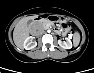
 CT scan axila section of the abdomen in 35 year old female patietn showing well defined mildly enhancing rounded soft tissue mass lesion seen in the retroperitoneal region on the right side causing displacement of the pancreas and other adjacent structures.
 Coronal reformatted image showing the mass lesion.
Axial T2 fat saturated MR image showing mixed intensity lesion.
The USG guided FNAC of the lesion proved to be paraganglioma.

Wednesday, August 1, 2012

Femoroacetabular impingement


Live ectopic pregnancy

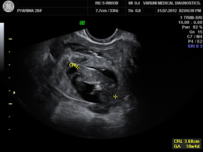
29 year old G2, P1 presents with 11 weeks of amenorrhea and pain abdomen. The urine pregnancy test was  positive.


Wednesday, February 8, 2012

Traumatic aortic dissection-CT Angiography


28 year old male patient came to emergency with road traffic accident, emergency thoracic CT angiography showed dissection of aortic arch distal to the left subclavian artery. Note the intimal flap (arrow) with true and false lumen (Type B dissection).

Discussion:

Up to 15% of all deaths following motor vehicle collisions are due to injury to the thoracic aorta. Many of these patients are dead at scene from complete aortic transection. Patients who survive to the emergency department usually have dissection, small tears or partial-thickness tears of the aortic wall with pseudoaneurysm formation.

Chest radiograph has a low sensitivity and has got high negetive predictive value (98%). Most sensitive indicator is abnormal appearing mediastinum, mediastinal widening of more than 8 cm at the level of aortic arch. Sensitivity of CT scan with the advent of MDCT is increased and is 97 to 100%. Intimal flap with true and false lumen are seen clearly.

Treatment is edovascular stent graft in type B dissection and surgical management if it is type A.

Isolated true anterior meningocele

Chest radiograph of 15 year old male showing mediastinal based large well defined mass lesion in the right upper hemithorax with defect in the vertebral body.


CT scan showing ventral defect in the vertebral body with herniation of meninges which is extending in to right hemithorax and displacing the trachea.

A spinal meningocele is a herniation of the meninges through a foramina or a defect in the vertebral column and is frequently located posteriorly in the lumbosacral area. 
An anterior spinal meningocele is rare and is generally described in the thoracic or sacral region. These frequently occur as a manifestation of generalized mesenchymal dysplasia such as neurofibromatosis type 1 (NF-1) or Marfan syndrome and rarely as an isolated defect.

CT and MR imaging are essential, not only for the diagnosis, but also for the depiction of its relationships to surrounding structures and the exclusion of other possible accompanying lesion such as neuroma in the setting of NF-1.

Thursday, January 12, 2012

Elbow Dislocation

32 Year old male with road traffic accident showing posterior dislocation of the elbow joint with mild proximal migration of the radius and ulna and small displaced chip fracture of the coronoid process.

MRI sagittal T2 images showing posterior dislocated elbow.


Discussion:
  • Elbow dislocation is the second most common major joint dislocation.
  • Dislocation is usually closed and posterior.
Mechanism:
  • Dislocations of elbow usually result from fall onto extended elbow. 
  • Anatomic morphology of semilunar notch may predispose to elbow dislocation.
  • Central angle of semilunar notch is significantly larger in group of pts who had dislocation of the elbow compared to normals.
Classification:
  • Classified according to direction of dislocation, namely posterior, posterolateral, posteromedial, lateral, medial, or divergent.
  • Simple - elbow dislocations without fracture.
  • Complex - dislocations with fracture. 
Simple dislocation:
  • Rupture of capsule, rupture of MCL, lateral ligaments, rupture of flexor pronator mass and less commonly, injury to brachialis muscle.
  • Rupture of brachial artery has been reported.
Complex dislocation:
  • Dislocation with radial head fracture is most common complex dislocation as in our case.
  • Fracture dislocation with MCL injury: radial head fracture & MCL Instability: 
  • Terrible Triad: (dislocation, cornoid process #, and radial head #).
Vascular injury: Closed dislocation rarely associated with vascular injury.

Neurologic injury:
  • Compartement syndrome.
  • Neuropraxia.
  • Ulnar nerve injury in 14%.
  • Entrapment of median nerve.

Wednesday, January 11, 2012

Phenytoin associated Cerebellar atrophy - MRI


28 year old female with chronic seizures on long term phenytoin therapy. MR examination of the brain showing diffuse atrophy of the both lobes of the cerebellum, vermis and the brainstem evidenced by prominent cerebellar foliae, pre pontine and CP angle cisterns.
Discussion:
Diffuse atrophy of the cerebellum refers to degeneration / reduction from a previously normal cerebellar volume. The term slightly differs from cerebellar hypoplasia meaning the cerebellum was not well formed to start with.
  • Diffuse atrophy can result from variety of causes
  • Normal ageing
  • Alcoholic cerebellar degeneration
  • Drugs (e.g phenytoin)
  • Multi system atrophy (MSA)
  • Ataxia telangiectasia 
  • Friedreich ataxia
  • Marinesco-Sjögren syndrome (MSS)
  • Fahr disease : atrophy not specific to cerebellum
  • Paraneoplastic cerebellar degeneration (PCD)
  • Olivopontocerebellar atrophy (OPCA)
  • Cerebrotenidinous xanthomatosis

Differential diagnoses
  • Cerebellar hypoplasia
  • Mega cisterna magna

Chondrosarcoma of Sternum - MRI

Chest Radiograph of the 70 year old female presenting with large mass growth in the sternal region showing significant widening of the superior mediastinum.

Axial T2 FS image showing large hyperintense mass lesion seen arising from the sternum with large retrosternal component causing mild mass effect on the mediastinal vessels.

T1 weighted axial image showing the lesion is hypo to isointense with retrosternal component.

Sagittal image showing large lesion in the sternal region and also extending superiorly in to the neck causing mass effect on the trachea.

Discussion:
  • It represent ~ 30% of primary malignant bone neoplasias, being the most frequent that of the anterior thoracic wall.
  • This tumor occurs more often between the third and fourth decade of life.
  • The males are more affected 
  • They are lobulated neoplasias that may grow to massive proportion and, consequently, may extend internally to the pleural space, or externally, invading muscle and adipose tissue of the thoracic wall. 
  • Microscopically, the findings vary from normal cartilage to obvious malignant modifications. The differentiation between chondroma and chondrosarcoma can be extremely difficult 
  • Palpable mass in thorax is the main symptom in approximately 80% of the patients with thoracic wall tumor. Of these, 60% present associated pain 
  • Respiratory failure and hemothorax are rare, and are present only in very large tumors 
Imaging:
  • Imaging exams may be useful to indicate the pathology and extent of the lesion; however, the definitive diagnostic requires a correlation between histology and radiology. 
  • Computerized tomography (CT) and magnetic resonance (MR) are good exams to characterize the tumor and its extension. 
  • CT is superior to MR to demonstrate calcifications, whereas MR is the choice to evaluate the tumor’s extension and its relationships with adjacent structures 
  • Thoracic wall chondrosarcomas typically grow slowly and relapse locally. If not treated, late metastasis will occur. 
Treatment:
  • Complete control of the primary neoplasia is the main determinant of survival. The purpose of the first surgery must be a wide resection, enough to prevent local recurrence.

Saturday, January 7, 2012

Cysticercosis (Muscular)- USG

Ultrasonography showing a well defined thick walled anechoic lesion with posterior acoustic enhancement seen in the medial aspect of the distal arm within the biceps brachi muscle. The lesion shows echogenic mural nodule attached to the posterior wall.

Transverse view of the same lesion showing cystic lesion with mural nodule

 Colour Doppler showing no evidence of colour flow in the lesion

Power Doppler showing no flow in the lesion.
The findings are consistent with Cysticercosis. Confirmed on HPR.

Uterine Arteriovenous Malformation - USG Doppler

Gray scale trans abdominal longitudinal view Ultrasonography of uterus in a 34 year old female showing multiple an-echoic areas within the myometrum of the anterior wall (arrow) displacing the thin linier endometrium (arrow head).

On colour Doppler there are multiple colour filled dilated vascular channels.

Power Doppler depicting the color flow within the lesion

Spectral wave form at the feeding artery showing high velocity and relatively high resistant arterial flow.

Spectral tracing in the draining vein showing venous flow. The findings are consistent with uterine arteriovenous fistula. Patient had history of recent medical termination of pregnancy with dilatation of curettage.

Discussion:

Uterine vascular malformations include:
  • Arteriovenous malformations (AVM) (especially arteriovenous fistulas).
  • True aneurysms.
  • Pseudoaneurysms. 
Uterine AVMs are rare in nonpregnant women.
It is otherwise called with several terms which are cavernous hemangioma, cirsoid aneurysm, racemose aneurysm, arteriovenous aneurysm, pulsatile angioma, and arteriovenous fistula.

Etiology:
AV fistulas are usually acquired and typically represent a single artery joining a single vein.
They result as a consequence of previous uterine trauma (eg, prior pelvic surgery, curettage), use of intrauterine contraceptive devices, pathologic pregnancy-related events, and previous treatment for gestational trophoblastic disease.

Clinical Features:
Vaginal bleeding- Most common.
Congestive heart failure - secondary to steal phenomenon - Less common.
Imaging findings:
USG:
  • Gray-scale US shows a normal-appearing endometrium, whereas the myometrium contains multiple hypoechoic or anechoic spaces.
  • At color Doppler US, these cystic spaces generate color signals in a mosaic pattern representing turbulent flow.
  • Spectral analysis of the arterial vessels within the lesion shows high-velocity flow with a low resistive index (approximately 0.51–0.65). Spectral analysis of venous flow demonstrates a similar pattern.
CT: CT angiography of the pelvic vessels may reveal the vascular malformation.

MRI:
Multiple dilated tubular channels showing flow related signal voids (hypointense) on T1 and T2 images seen in myometrium with normal endometrium or displaced endometrium. The flow relted signal void channels may or may not extend in to the parametrium.

Treatment:
  • Acute treatment - Hemodynamic stabilization and management of active bleeding. 
  • Occlusion with a Foley catheter bulb may be effective. 
  • If fertility is not an issue, hysterectomy is the treatment of choice 
  • They may be treated successfully with intra-arterial embolization

*******

Monday, December 5, 2011

Ovarian Dermoid - CT

CT scan axial section in a 28 Year old female with pain abdomen since 2 years showing well defined predominantly fat density round mass lesion seen in the pelvis in the mid line and  towards right side with areas of calcification (Rokintasky protruberence), soft tissue and fluid components in the dependent part. Features are consistent with dermoid. 

Discussion:
A term dermoid is used now a days instead of teratomas which derived from more than one germ layer. 
The contents of the dermoid include hair, teeth, fat, skin, muscle, endocrine tissue and ectodermal tissue predominates among these.
  • Mature cystic teratomas are commonly referred as dermoid cysts.
  • They are most common ovarian neoplasm.
  • Mature cystic teratomas of the ovary is invariably benign. Rarely (1-3%) my undergo malignant. Prognosis is poor for such tumors.

Age: Can occur at any age. More common during reproductive age (16-55) and peak at 20-40. 50% of adnexal neoplasms are dermoid during adolescence. 

Location:  Most commonly ovary, bilateral in 8-25%.
Other locations: Mediastinum, retroperitoneum, cervical region, brain

Clinical findings: Frequently incidentally detected.
  • Abdominal pain as in our patient.
  • Mass
  • Abnormal uterine bleeding.
  • Back pain.
  • Bladder and GI symptoms.
Imaging findings:

Radiograph:
  • A soft tissue mass if it is large.
  • A fat containing mass of fat density - the density of mass is lower than surrounding tissues.
  • Characteristic calcification -Pop cord calcification or rim like or tooth or other bone (clavicle).
Ultrasonography:

  • It has been reported as 98% of positive predictive value for ovarian dermoids.
  • Compled mass with echogenic components.
  • Mass is echogenic most of the times and producing "dirty acoustic shadowing"
  • It may be purely cystic in 9-15% or purely solid in 10-31% cases.
CT scan:

  • It demonstrates mass having fat, fluid and calcification (Tooth) as its contents.
  • Rim like calcification, Tooth or other bone (clavicle) -Rokintasky protruberence.
  • Fat-fluid levels may be seen.
MRI:
  • The lesion shows hyperintense fat on T1 with hypointense fluid.
  • Hyperintense on T2.
  • Fat appears hypointense (suppressed) on STIR or T2 fat sat images.
Treatment:
  • Surgical removal - May have risk of chemical peritonitis if ruptured.
Complications:
  • Torsion.
  • Rupture.
  • Infection.
  • Autoimmune hemolytic anemia - very rare.
Differential diagnosis:
  • Benign or malignant ovarian tumor.
  • Endometrioma.
  • TO Abscess.
  • Pedunculated uterine fibroid.
  • Hydrosalpinx.
  • Ectopic kidney.
  • Pelvic kidney.

    Monday, November 14, 2011

    Carcinoma lung on the background of ILD - CT

    Radiograph of 65 year old male patient who is know case of Usual interstitial pneumonitis (UIP) showing bilateral reticular shadows (arrow heads) with well deifned soft tissue density lesion in the left upper lobe perihiar region. 


    CT scan at the level of arch of aorta showing heterogenpusly enhancing lesion in the left upper lobe (arrow) and HRCT lung window showing reticular shadows with honey combing (arrow head). CT guided biopsy of the lesion showed non small cell bronchogenic carcinoma. 

    Discussion:

    Studies have showed that the Usual interstitial pneumonia (UIP), or idiopathic pulmonary fibrosis (IPF), has been considered to be associated with a high risk for lung carcinoma. It is more common in males probably due to smoking. 

    The nodule or mass in a patient with UIP should be evaluated properly. CT and CT-PET are the preferred modalities. If requred CT guidded biopsy has to be done to confirm the diagnosis. As small as 1 cm nodule can be sampled with CT guidence. 

    Most of the carcinomas will be peripheral in patients with UIP unlike our case. The diseased lung (UIP) surrounding the nodule/mass can cause pneumothorax during biopsy should be taken care meticulously.

    Tracheal trifurcation-CT and Virtual bronchoscopy


    A 2 year old boy admitted with recurrent respiratory tract infection for evaluation of active chest infection. Multislice computed tomography of chest was done which showed no evidence of active infection, but there was an incidental finding of tracheal trifurcation (volume rendered reconstruction showed two bronchi [long white arrow and arrow head] arising from the trachea on right side and left main bronchus [short arrow], and on virtual bronchoscopy inset two bronchi [long white arrow and arrow head] and left main bronchus [short arrow]).

    Discussion:

    Tracheal trifurcation is an extremely rare anomaly in which the trachea divides into three main stem bronchi rather than two, with two main bronchi supplying the right lung and one supplying the left lung.

    Patients usually are asymptomatic and it is detected incidentally on imaging. Some patients may present with recurrent respiratory tract infection and may progress to bronchiectasis and chronic bronchitis.

    It is important to identify this anatomical variant in patients requiring tracheal intubation.

    Search This Blog