Showing posts with label Neuroradiology. Show all posts

Wednesday, February 8, 2012

Isolated true anterior meningocele

Chest radiograph of 15 year old male showing mediastinal based large well defined mass lesion in the right upper hemithorax with defect in the vertebral body.


CT scan showing ventral defect in the vertebral body with herniation of meninges which is extending in to right hemithorax and displacing the trachea.

A spinal meningocele is a herniation of the meninges through a foramina or a defect in the vertebral column and is frequently located posteriorly in the lumbosacral area. 
An anterior spinal meningocele is rare and is generally described in the thoracic or sacral region. These frequently occur as a manifestation of generalized mesenchymal dysplasia such as neurofibromatosis type 1 (NF-1) or Marfan syndrome and rarely as an isolated defect.

CT and MR imaging are essential, not only for the diagnosis, but also for the depiction of its relationships to surrounding structures and the exclusion of other possible accompanying lesion such as neuroma in the setting of NF-1.

Wednesday, January 11, 2012

Phenytoin associated Cerebellar atrophy - MRI


28 year old female with chronic seizures on long term phenytoin therapy. MR examination of the brain showing diffuse atrophy of the both lobes of the cerebellum, vermis and the brainstem evidenced by prominent cerebellar foliae, pre pontine and CP angle cisterns.
Discussion:
Diffuse atrophy of the cerebellum refers to degeneration / reduction from a previously normal cerebellar volume. The term slightly differs from cerebellar hypoplasia meaning the cerebellum was not well formed to start with.
  • Diffuse atrophy can result from variety of causes
  • Normal ageing
  • Alcoholic cerebellar degeneration
  • Drugs (e.g phenytoin)
  • Multi system atrophy (MSA)
  • Ataxia telangiectasia 
  • Friedreich ataxia
  • Marinesco-Sjögren syndrome (MSS)
  • Fahr disease : atrophy not specific to cerebellum
  • Paraneoplastic cerebellar degeneration (PCD)
  • Olivopontocerebellar atrophy (OPCA)
  • Cerebrotenidinous xanthomatosis

Differential diagnoses
  • Cerebellar hypoplasia
  • Mega cisterna magna

Monday, October 17, 2011

Unusual migration of proximal tip of ventriculoperitoneal shunt - CT

26 year old male patient with post traumatic hydrocephalus treated with ventriculoperitoneal shunt axial CT sectio at hte level of third ventricle showing tubular hyperdense structure (arrow) suggestive of VP shunt.

Axial section at the level of basal cisterns showing the VP shunt catheter in the suprasellar structures.

Sagittal reformatted image showing the catheter tip is situated at the roof of the sella.

Discussion:
  • There are many incidents of unusual migration of the shunt tip in to various parts of the body. 
  • Usually it is the distal or the peritoneal catheter that migrates after breakage or disconnection into many locations such as the scalp, heart, anus/rectum, urethral, knee, umbilicus, chest, pleural cavity, inguinal canal and scrotum.
  • Complete migration of the shunt in to the ventricles have been reported.
  • This might be the first case of shunt migration in to the suprasellar region as far as my knowledge is concerned.

Tuesday, September 27, 2011

Calvarial thickening - MRI

T1 weighted axial image in 24 year old female with thalasemia showing diffuse thickening of the calvarium.

T2 weighted axial image showing calvarial thickening.

FLAIR axial image showing calvarial thickening.

Discussion:
Differential diagnosis of calvarial thickening:
  1. Various severe anemias - bone marrow expansion.
  2. Pagets disease.
  3. Hyperparathyroidism.
  4. Osteopetrosis.
  5. Chronic Dilatin therapy.
  6. Acromegaly.
  7. Some rare genetic diseases - Camurati-Engelmann’s disease, frontometaphyseal dysplasia and   craniodiaphysial dysplasia.

Tuesday, August 23, 2011

Gray matter heterotopia - MRI


T1, T2 and T1 inversion recovery axial image in 16 year old female patient with seizures showing smooth, nodular area, isointense to grey matter seen on all the sequences and present all along the margin of the bilateral lateral ventricles (subependymal regions) consistent with gray matter heterotopia.The lesion is better depicted in T1 inversion recvoery image.

Discussion:
Definition: It is a type of cortical dysplasia (neuronal migrational anomaly) where in clumps of normal grey matter being located in the wrong part of the brain.
Clinical features: Asymptomatic to profound seizures and it is usually detected incidentally on brain MRI. Various manifestations include severe seizure disorder, loss of motor skills, and mental retardation. Fatalities are practically unknown, other than the death of unborn male fetuses with a specific genetic defect.
Types of Heterotopia:
  1. Periventricular or subependymal: It can be seen as small single node or a large number of nodes, can exist on either or both sides of the brain at any point along the higher ventricle margins, can be small or large, single or multiple, and can form a small node or a large wavy or curved mass
  2. Focal Subcortical: form as distinct nodes in the white matter, "focal" indicating specific area.
  3. Band Form: "Band" heterotopia form in the white matter beneath the cortex, but the gray matter is more diffuse and is symmetric between the hemispheres.
Imaging: CT or MRI
CT scan: The density will be similar to that of the gray matter.
MRI: The signal intensity will be similar to gray matter in all the sequences. T1 inversion recovery sequence will better depict the lesion.

Monday, July 4, 2011

Neurocysticercosis starry sky appearance - MRI

Contrast enhanced T1 weighted axial image showing multiple nodular and ring enhancing lesions in bilateral cerebral hemispheres giving starry sky appearance.

Contrast enhanced T1 weighted coronal image showing multiple nodular and ring enhancing lesions in bilateral cerebral hemispheres.

Colloid cyst - MRI

Axial non enhanced T1 magnetic resonance image showing a bright mass in the roof of third ventricle (foramen of Monro).

Axial non enhanced T2 magnetic resonance image showing a hypointense mass in the roof of third ventricle.

Sagittal non enhanced T1-weighted magnetic resonance image. This image demonstrates a round area of increased signal intensity lesion in the anterosuperior portion of the third ventricle.

Discussion:
Colloid cysts are benign, congenital epithelium-lined cysts that almost always arise in the anterior third entricle. However, rare reports describe cysts in other locations. The cysts are believed to derive from either primitive neuroepithelium of the tela choroidea or from endoderm.
Patient may present with intermittent head ache. Rarely m
ay cause sudden death .

CT scan:
  • Rounded small mass with high attenuation at the anterior third ventricle - due to proteinacious material.
  • Rarely the lesion is isoattenuating or hypoattenuating relative to brain parenchyma.
  • Intermittent hydrocephalus - colloid cyst act as ball valve.
MRI:
  • Usually found at the foramen of Monro just posterior to the fornices in the anterosuperior third ventricle.
  • MR signal intensities are variable: Variation due to contents of the cyst.
  • Commonly hyperintense on T1 and iso to hypointense on T2.
  • Colloid cysts do not have intrinsic enhancement; the presence of enhancement suggests a solid tumor. Peripheral enhancement may be present because of vascularity in the outer wall.

Tuesday, June 28, 2011

Hot cross bun sign - Multiple system atrophy


Transverse and sagittal T2-weighted MRimage of the brain of a patient with multiple system atrophy (MSA) of the cerebellar-predominant subtype (MSA-c) shows the hot cross bun sign as a cruciform hyperintensity
in an atrophied pons. Cerebellum and middle cerebellar peduncles are also atrophied.

Discussion:
Multiple system atrophy is a rare neurological disorder characterized by a combination of parkinsonism, cerebellar and pyramidal signs, and autonomic dysfunction. The term "Multiple System Atrophy" is synonymous with striatonigral degeneration (SND) when Parkinsonism predominates, olivopontocerebellar atrophy (OPCA) when cerebellar signs predominate, and Shy-Drager syndrome when autonomic failure is dominant. The incidence (new case per 100,000 person years) for ages 50 to 99 years is 3.0 (Bower et al, 1997), or about half as frequent as it's close relative, progressive supranuclear palsy (PSP). The mean age of onset is 54.

Pathophysiology: There is neuronal loss and gliosis in the inferior olives, pons, cerebellum, substantia nigra, locus ceruleus, striatum and the intermediolateral column of the spinal cord.
Similar pattern of neuronal loss is seen in a patient with parkinsonism, the neuronal loss being secondary to presumed vasculitis, and proposed that the sign may reflect wallerian degeneration of transverse pontocerebellar fibers secondary to vasculitic infarction.\


MRI - On T2-weighted MR images, a hyperintense rim at the putaminal edge, putaminal atrophy, and intrinsic signal intensity change are seen. In MSA-c, the changes predominantly affect the infratentorial structures. On T2-weighted MR images, atrophy and increased signal intensity within the pons, cerebellum, and middle cerebellar peduncles are seen.


References
1. Schrag A, Kingsley D, Phatouros C, et al. Clinical usefulness of magnetic resonance imaging in multiple system atrophy. J Neurol Neurosurg Psychiatry 1998;65:65–71.
2. Muqit MM, Mort D, Miskiel KA, Shakir RA. “Hot cross bun” sign in a patient with parkinsonism secondary to presumed vasculitis. J Neurol Neurosurg Psychiatry 2001;71:565–566.

Monday, April 11, 2011

Spondylolisthesis-MRI

T2 weighted sagittal image of the lumbar spine in a 44 year old male patient showing grade I spondylolisthesis at L3-4 (anterolisthesis of L3 over L4) with disc herniation. No traversing nerve roots compression. 

T2 weighted sagittal image of the lumbar spine right lateral most image and the left lateral most images showing exiting nerve root compression (arrow) and break in the pars inter articularis suggesting spondylolysis.

Axial T2 weighted image at L3-4 showing nerve root compression bilaterally.

Discussion:

Spondylolisthesis is officially categorized into five different types by the Wiltse classification system: Dysplastic, Isthmic, Degenerative, Traumatic, and Pathologic.

Grades:
  • Grade 1 is 0–25%
  • Grade 2 is 25–50%
  • Grade 3 is 50–75%
  • Grade 4 is 75–100%
  • Over 100% is Spondyloptosis, when the vertebra completely falls off the supporting vertebra.

Disc extrusion - MRI


T2 weighted sagittal MR images of the lumbar spine showing central disc extrusion with cranial migration.

Discussion:
A disk is classified as an extrusion if any distance between the edges of the disk material beyond the disk space is greater than the distance between the edges of the base measured in the same plane.

Extruded disk material that has no continuity with the disk of origin may be further characterized as sequestrated. A sequestrated disk is a subtype of extruded disk. By definition, a sequestrated disk can never be classified as a protruded disk. Disk material that is displaced away from the site of extrusion, regardless of the presence or absence of continuity, may be called migrated, a term that is useful for interpreting images because it is often impossible to show continuity on imaging.

Friday, April 8, 2011

Epidermoid

T2 weighted axial image of the 22 year old gentle man showing well defined hyperintense mass lesion in the basal cisterns on right side causing significant mass effect on the temporal horn of ipsilateral lateral ventricle.

T1 weighted axial image showing the lesion is hypointense to gray matter.


FLAIR axial image the lesion is showing mixed signal intensity predominantly hypointense.

Coronal T2 WI showing the lesion.

Axial B1000 Diffusion weighted image showing moderate restriction.

ADC image showing hypointense mass, which differentiates epidermoid from arachnoid cyst where the lesion will be hyperintense on ADC images.

Discussion:

Epidermoid cysts (sebaceous cysts) are benign congenital lesions of ectodermal origin. They account for approximately 1% of all intracranial tumors. Although these lesions are congenital, patients are usually not symptomatic until they are aged 20-40 years.

CT scan:

  • Low attenuation - similar to CSF density.
  • No enhancement.
  • Calcification in 15-20%.
  • Rarely they can be hyperdense secondary to high density.
MRI:
  • Isointense to CSF on T1 and T2 weighted images. T1 signal intensity varies with the amount of lipid content.
  • May be hyperintense on FLAIR images.
  • High signal on diffusion weighted images which differentiate from the low signal arachnoid cyst.
  • Minimal rim enhancement on post contrast images.

Arachnoid cyst
  • CSF filled/signal
  • Middle cranial fossa (MC) > cerebral convexities, basal cisterns, retrocerebellar region
  • Hypogenesis of adjacent brain
  • No enhancement
  • Low on DWI (like ventricle).

Monday, January 31, 2011

Neurocysticercosis - MRI


MRI Brain of 26 year old male presented with seizures, T1 image showing well defined small hypointense lesion in the right frontal cortical region, which is showing hyperintense on T2 image. CISS 3D - heavily weighted T2 images (bottom row) showing fluid intensity lesion with small hypointense focus. The findings are consistent with neurocysticercosis.


Syrinx - MRI


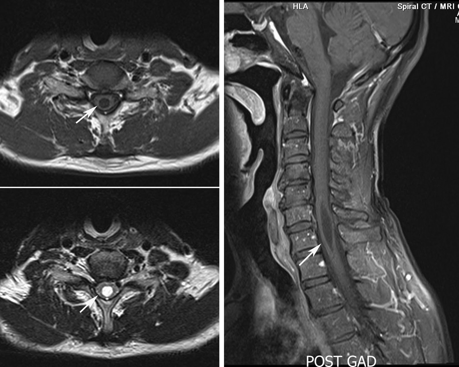
Cervical spine MRI in a 54 year old male patient showing focal dilatation of the central spinal canal with no enhancing focal lesion within the cord. The features are consistent with the focal syrinx.

Discussion:
Hydromyelia: An accumulation of cerebrospinal fluid (CSF) may lead to simple distention of the central canal of the spinal cord lined by ependymal cells.
Syringomyelia: an accumulation of CSF may dissect into the surrounding white matter to form a paracentral cavity, which is not lined by ependyma.
syringohydromyelia: Combination of both which is seen in most of the cases.

I - In 1973, Barnett et al classified syringohydromyelic cavities into 5 types:
  1. Communicating (with the subarachnoid space, usually at the level of the obex at the inferior aspect of the fourth ventricle)
  2. Posttraumatic
  3. Tumor-related
  4. Arachnoiditis-related
  5. Idiopathic.
II - Milhorat et al, the intramedullary cavities were classified into communicating, noncommunicating, and atrophic types.

III -  Noncommunicating syringes are subdivided into 6 types:
  1. Chiari II malformation with hydrocephalus
  2. Chiari I malformation without hydrocephalus
  3. Extramedullary compressive lesions at the craniocervical junction or along the length of the spinal canal
  4. Spinal cord trauma
  5. Intramedullary tumors and intraperimedullary infections
  6. Multiple sclerosis

Thursday, January 13, 2011

HIV Encephalopathy - MRI


MRI axial fluid attenuation inversion recovery (FLAIR) images in 48 year old male with HIV showing generalised cerebral atrophy with diffuse white matter hyperintenisty signal change seen in the bilateral periventricular frontal and paroeto-occipital regions consistent with HIV encephalopathy.

Discussion:
Clinically it is usually presents with progressive dementia, initially cognitive impairment and later accompanied by motor symptoms like gait disturbance and tremor. Collectively this is called as AIDS dementia complex.
Histopathology may reveal subacute encephalitis, cerebral atrophy and demyelination.

CT - The sole finding in most of the patients may be cerebral atrophy. In progressive advanced patient may see periventricular confluent white matter hypodensities. 

MRI- Cerebral atrophy, classical appearance of confluent, bilateral and symmetrical white matter lesions seen as diffuse white matter change in the periventricular regions and centrum semiovale, with relativesparing of the subcortical white matter and posterior fossa structures. These lesions shows hyperintense on T1 and iso to hypointense on T1 images. There will be no contrast enhancement or mass effect.
MR Spectroscopy shows decreased N Acetyl Aspartate and increased choline peaks and changes in the glutamate and glutamine levels. 

Sunday, January 3, 2010

Primitive Neuroectodermal Tumor (PNET) of Skull vault : CT and MRI

4 year old female child came with history of mass in the right side of the head in parietal region. CT scan shows large heterogenously enhancing soft tissue mass lesion seen in both intra and extracranial regions, the intracranial component is extradural causing mass effect on the right cerebellar hemisphere. There is bone erosion of right parietal bone.

MRI of the same patient T2 WI showing similar findings with large extra and intracranial components. There is complete involvement of the right parietal bone. Biopsy proved to be PNET.

Primitive Neuroectodermal Tumor (PNET) is a soft tissue sarcoma classically described under small round blue cell tumors (SRBCT). Synonyms include : PNET, Peripheral neuroepithelioma, Peripheal neuroblastoma, Askin tumor (Thoracopulmonary PNET) and Extraosseous Ewing's sarcoma. The incidence is 1% of the sarcomas. They often (75%) occure in the age of less than 35 years. Most investigators now believe that Ewing's sarcoma and PNET are different morphological expressions of one tumor type. In general, Ewing's sarcoma arises within the bone while PNET arises within soft tissues. PNET arising from the dura or skull vault is very rare and in this case it is diffucult to predict the origin of the tumor.Radiograph is often the first investigation to be done. CT and MRI are the investigations of choice to see for the extent of the lesion. MRI is preferred over CT scan. These can be used to look for the response to chemo and radiotherapy.Here is the link to the PNET at various sites:

Sunday, December 27, 2009

Methylmalonic acidaemia (MMA)

3 year old female child came with developmental delay, lethargy, seizures and hypoglycemia, MRI brain T2 weighted image at the level of basal ganglia showing bilateral symmetrical hyperintensities in globi pallidi.

MMA encompasses a heterogeneous group of disorders characterized by accumulation of methylmalonic acid and its by-products in biological fluids. These disorders are due to a deficiency of the adenosylcobalamin-dependent enzyme methylmalonyl-CoA mutase (apoenzyme deficiency), a defect in intracellular cobalamin metabolism (coenzyme deficiency), transcobalamin II deficiency, intrinsic factor deficiency, or dietary cobalamin deficiency, which is found in vegetarians. The incidence, as derived form a neonatal screening program, is one in 48,000 live births (6). The actual occurrence, however, is estimated to be one in 25,000 live births.
CT and MR imaging of the brain typically reveal atrophy, delay in myelination, and abnormalities in the basal ganglia, predominantly in the globi pallidi. The globi pallidi are particularly sensitive to mitochondrial dysfunction, and are thus prime targets for injury. MR Spectroscopy may show elevated lactate levels either in CSF or in brain parenchyma i.e regions of infarcts. Differential diagnosis for bilateral globi pallidi lesions are HIE (hypoxic ischemic encephalopathy), kernicterus, mitochondrial encephalopathy etc.

Search This Blog