Showing posts with label Head and Neck. Show all posts

Tuesday, September 27, 2011

Traumatic optic nerve compression - CT

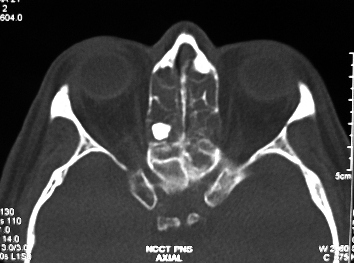
Axial CT section through the optic nerves in a 59 year old male with trauma showing fracture in the posterior part of the lateral wall of the right orbit and the fracture fragment is causing compression on the optic nerve at the optic canal (arrow) and note the normal wide optic canal on left side (arrow head).

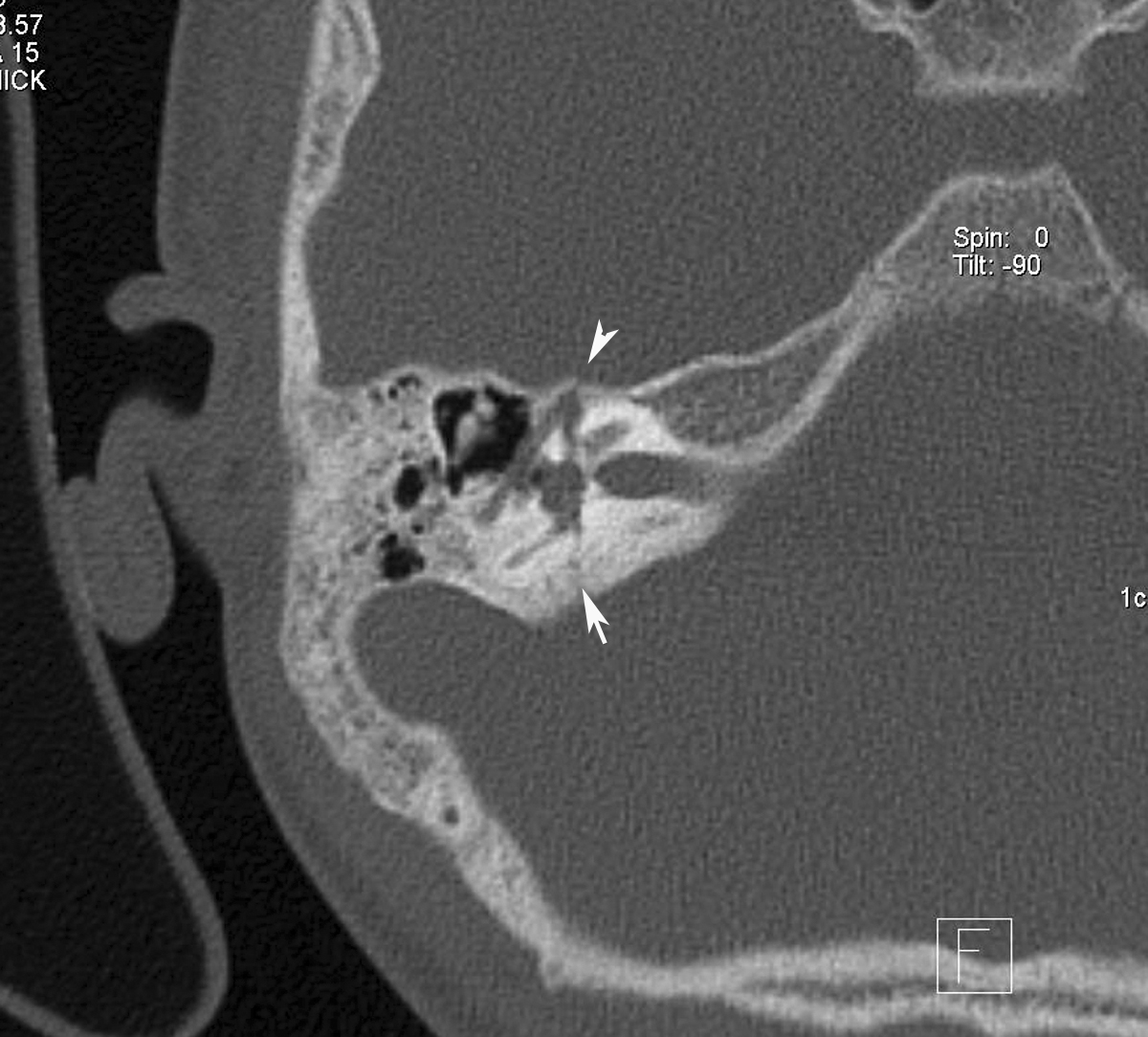
Coronal CT scan bone window showing the bony fragment compressing the optic nerve and narrowing the optic foramen (arrow) and note the normal foramen on left side (arrow head).

Sagittal image showing the bony fragment compressing on the optic nerve.
Discussion:
  • Traumatic optic neuropathy is a devastating potential complication of closed head injury.
  • The hallmark of an optic neuropathy, traumatic or otherwise, is a loss of visual function, which can manifest by subnormal visual acuity, visual field loss, or color vision dysfunction.
  • Vision loss associated with traumatic optic neuropathy can be partial or complete and temporary or permanent.
  • Best diagnostic test is Thin-slice CT scan of the nose, sinuses, and orbits
  • Fractures through the optic canal can be best depicted with thin-section CT scanning (eg, 1.5-mm cuts with 1-mm intervals).
  • CT scanning provides adequate imaging of orbital soft tissue and is better than MRI at delineating bony defects. 
  • It provides an intraoperative road map for the surgeon in patients who require surgical decompression and can be used for image guided endoscopic surgery.
  • Treatment is observation, steroids and optic nerve decompression. The decision for surgical decompression should still be based primarily on the clinical examination findings.

Saturday, September 10, 2011

Temporal bone fracture with facial nerve injury - HRCT

HRCT of right temporal bone axial image showing transverse temporal bone fracture (arrow) and the fracture line passing through the first anterior genu of the facial nerve canal (arrow head).

Subsequent section of the HRCT right temporal bone showing the fracture passing through the facial nerve canal (arrow head).

Discussion: Temporal bone fractures classically are described with reference to the long axis of the petrous bone.
Classification:
1. Longitudinal (parallel to the axis).
2. Transverse (perpendicular to the axis).
3. Oblique/Mixed- Common.


Longitudinal fracture: The fracture line of force runs roughly from lateral to medial. A fracture line may extend through the facial nerve canal, thereby damaging the facial nerve. Associated injury, such as transection or intraneural hemorrhage, may cause facial nerve paralysis, as can damage from displaced bone fragments.


Transverse fracture: result from trauma to the occiput or cranial-cervical junction, with the line of force running roughly anterior to posterior.A fracture passing through the vestibulocochlear apparatus can cause sensorineural hearing loss and equilibrium disorders. Transverse fractures also commonly injure CN VII, because their path often takes them close to the nerve's labyrinthine segment.

Mixed fractures: Oblique (or mixed) fracture patterns, which extend both longitudinally and transversely, are common, and some case series report that these occur more often than do isolated transverse or longitudinal fractures.

Radiology:
Radiography: Plain film radiographs of the skull may show opacified mastoid air cells, intracranial air, or, rarely, a lucency (fracture line).

HRCT: can demonstrate a lucency through the temporal bone. Involvement of the middle ear, petrous bone, otic capsule, and facial nerve canal are the primary determinants of prognosis.


MRI: may demonstrate fluid (high signal on T2-weighted images) in the middle ear and mastoid air cells. T1-weighted images may reveal a bright signal in the labyrinth or middle ear, consistent with hemorrhage.

Sunday, July 3, 2011

Ivory Osteoma

Coronal CT paranasal sinuses showing well defined rounded hyperdense focus seen in the right ethmoid air cell suggestive of ivory osteoma
All the sinuses shows soft tissue opacification including the nasal cavity due to allergic fungal rhinosinusitis.

Axial CT section showing the Ivory osteoma in the posterior ethmoid air cell

Discussion:
Most common tumor of the paranasal sinuses and frequently seen in the frontal and ethmoid sinuses. It is benign tumor of membranous bone consisting of dense, compact bone and majority of of them are discovered serendipitously.
In the skull, they usually arise from the outer table. Rarely, large osteoma in the frontal or ethmoid region may displace globe forward and cause proptosis.
Obstruction of a sinus ostium may lead to infection or formation of a mucocele. Very rarely, an osteoma may erode through the dura leading to cerebrospinal fluid rhinorrhea or intracranial infection.

Imaging findings:
Well-circumscribed, sharply-marginated round and very dense lesions usually less than 2 cm in size. Multiple paranasal osteomas are found in Gardner’s syndrome.
Multiple osteoma of the mandible and maxilla, along with the frontal, sphenoid and ethmoid sinuses, rarely the long bones or phalanges
Association between colonic polyps with a predilection to malignant degeneration.

Friday, April 1, 2011

Frontoethmoidal Encephalocele

13 Year old female with hard swelling in the fore head. Volume rendering colour coded image of the face showing fromtoethmoidal encephalocele.

Volume rending image with bone algorithm showing bony prominence in the frontoethmoidal region. 

Cropped volume rendered image to show the defect in the frontoethmoid region

Oblique coronal reformatted bone window image showing the defect. 

Sagittal reformatted bone window image showing the defect clearly.

Coronal T2 fat saturated MR image showing herniation of the meninges through the above showed defect.

Discussion:

Encephalocele can be a congenital or an acquired abnormality of the brain in which intracranial contents including meninges, CSF, and brain tissue herniate through a skull defect. Congenital encephaloceles occur when the mesodermal layer between the neural tube and the ectoderm fails to develop and the anterior neuropore remains open. Anterior encephaloceles can also occur after trauma or surgery. Brain pulsations are presumed to push brain tissue through the defect. Patients are prone to recurrent episodes of meningitis. In addition, visual acuity and hypothalamic function may be affected. Clinical presentation includes a nasopharyngeal mass, which may enlarge with Valsalva's maneuver.

Differential considerations include a tumor traversing the cribriform plate, granuloma or esthesioneuroblastoma.

Thursday, September 30, 2010

Cholesteatoma- HRCT

Non contrasted temporal bone CT reveals a soft tissue mass in the right middle ear and Prussak’s space with associated erosions of the scutum, epitympanum walls, lateral semicircular canal, tegmen tympani and middle ear ossicles. It is also extending in to the mastoid air cells.
Discussion:
  • An acquired cholesteatoma is a collection of exfoliated squamous epithelium and debris.
  • Cholesteatomas often begin at the pars flaccida of the tympanic membrane and grow in Prussak's space of the temporal bone and produce inflammatory reaction.
  • Cholesteatomas typically occur in the setting of chronic dysfunction of the Eustachian tubes and recurrent otitis media.
  • Cholesteatomas can be as small as a couple of millimeters in size or can grow to fill the entire middle ear.
  • Erosions of the scutum and ossicles are commonly seen.
  • The presence of bony erosions or expansion are strong support of cholesteatoma over chronic otomastoiditis.
  • Acquired cholesteatomas occur in children and adults.
  • Most common presenting symptoms include ear discharge, hearing loss, ear pain and vertigo.
  • Without treatment cholesteatomas will progressively increase in size.
  • Complications include: Hearing loss, CN 7 palsy, venous sinus thrombosis, semicircular canal fistulas, and intracranial invasion.
  • Early surgical intervention usually results in complete eradication and preservation of hearing.
Differential diagnosis:
  1. Acquired cholesteatoma
  2. Chronic otomastoiditis
  3. Middle ear cholesterol granuloma
  4. Glomus tympanicum paraganglioma

Search This Blog