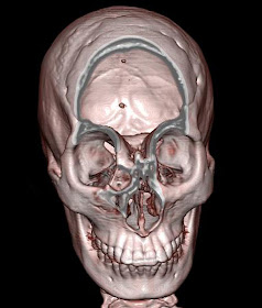
13 Year old female with hard swelling in the fore head. Volume rendering colour coded image of the face showing fromtoethmoidal encephalocele.
Volume rending image with bone algorithm showing bony prominence in the frontoethmoidal region.
Cropped volume rendered image to show the defect in the frontoethmoid region
Oblique coronal reformatted bone window image showing the defect.
Sagittal reformatted bone window image showing the defect clearly.
Coronal T2 fat saturated MR image showing herniation of the meninges through the above showed defect.
Discussion:
Encephalocele can be a congenital or an acquired abnormality of the brain in which intracranial contents including meninges, CSF, and brain tissue herniate through a skull defect. Congenital encephaloceles occur when the mesodermal layer between the neural tube and the ectoderm fails to develop and the anterior neuropore remains open. Anterior encephaloceles can also occur after trauma or surgery. Brain pulsations are presumed to push brain tissue through the defect. Patients are prone to recurrent episodes of meningitis. In addition, visual acuity and hypothalamic function may be affected. Clinical presentation includes a nasopharyngeal mass, which may enlarge with Valsalva's maneuver.
Differential considerations include a tumor traversing the cribriform plate, granuloma or esthesioneuroblastoma.


No comments:
Post a Comment