Monday, December 5, 2011

Ovarian Dermoid - CT

CT scan axial section in a 28 Year old female with pain abdomen since 2 years showing well defined predominantly fat density round mass lesion seen in the pelvis in the mid line and  towards right side with areas of calcification (Rokintasky protruberence), soft tissue and fluid components in the dependent part. Features are consistent with dermoid. 

Discussion:
A term dermoid is used now a days instead of teratomas which derived from more than one germ layer. 
The contents of the dermoid include hair, teeth, fat, skin, muscle, endocrine tissue and ectodermal tissue predominates among these.
  • Mature cystic teratomas are commonly referred as dermoid cysts.
  • They are most common ovarian neoplasm.
  • Mature cystic teratomas of the ovary is invariably benign. Rarely (1-3%) my undergo malignant. Prognosis is poor for such tumors.

Age: Can occur at any age. More common during reproductive age (16-55) and peak at 20-40. 50% of adnexal neoplasms are dermoid during adolescence. 

Location:  Most commonly ovary, bilateral in 8-25%.
Other locations: Mediastinum, retroperitoneum, cervical region, brain

Clinical findings: Frequently incidentally detected.
  • Abdominal pain as in our patient.
  • Mass
  • Abnormal uterine bleeding.
  • Back pain.
  • Bladder and GI symptoms.
Imaging findings:

Radiograph:
  • A soft tissue mass if it is large.
  • A fat containing mass of fat density - the density of mass is lower than surrounding tissues.
  • Characteristic calcification -Pop cord calcification or rim like or tooth or other bone (clavicle).
Ultrasonography:

  • It has been reported as 98% of positive predictive value for ovarian dermoids.
  • Compled mass with echogenic components.
  • Mass is echogenic most of the times and producing "dirty acoustic shadowing"
  • It may be purely cystic in 9-15% or purely solid in 10-31% cases.
CT scan:

  • It demonstrates mass having fat, fluid and calcification (Tooth) as its contents.
  • Rim like calcification, Tooth or other bone (clavicle) -Rokintasky protruberence.
  • Fat-fluid levels may be seen.
MRI:
  • The lesion shows hyperintense fat on T1 with hypointense fluid.
  • Hyperintense on T2.
  • Fat appears hypointense (suppressed) on STIR or T2 fat sat images.
Treatment:
  • Surgical removal - May have risk of chemical peritonitis if ruptured.
Complications:
  • Torsion.
  • Rupture.
  • Infection.
  • Autoimmune hemolytic anemia - very rare.
Differential diagnosis:
  • Benign or malignant ovarian tumor.
  • Endometrioma.
  • TO Abscess.
  • Pedunculated uterine fibroid.
  • Hydrosalpinx.
  • Ectopic kidney.
  • Pelvic kidney.

    Monday, November 14, 2011

    Carcinoma lung on the background of ILD - CT

    Radiograph of 65 year old male patient who is know case of Usual interstitial pneumonitis (UIP) showing bilateral reticular shadows (arrow heads) with well deifned soft tissue density lesion in the left upper lobe perihiar region. 


    CT scan at the level of arch of aorta showing heterogenpusly enhancing lesion in the left upper lobe (arrow) and HRCT lung window showing reticular shadows with honey combing (arrow head). CT guided biopsy of the lesion showed non small cell bronchogenic carcinoma. 

    Discussion:

    Studies have showed that the Usual interstitial pneumonia (UIP), or idiopathic pulmonary fibrosis (IPF), has been considered to be associated with a high risk for lung carcinoma. It is more common in males probably due to smoking. 

    The nodule or mass in a patient with UIP should be evaluated properly. CT and CT-PET are the preferred modalities. If requred CT guidded biopsy has to be done to confirm the diagnosis. As small as 1 cm nodule can be sampled with CT guidence. 

    Most of the carcinomas will be peripheral in patients with UIP unlike our case. The diseased lung (UIP) surrounding the nodule/mass can cause pneumothorax during biopsy should be taken care meticulously.

    Tracheal trifurcation-CT and Virtual bronchoscopy


    A 2 year old boy admitted with recurrent respiratory tract infection for evaluation of active chest infection. Multislice computed tomography of chest was done which showed no evidence of active infection, but there was an incidental finding of tracheal trifurcation (volume rendered reconstruction showed two bronchi [long white arrow and arrow head] arising from the trachea on right side and left main bronchus [short arrow], and on virtual bronchoscopy inset two bronchi [long white arrow and arrow head] and left main bronchus [short arrow]).

    Discussion:

    Tracheal trifurcation is an extremely rare anomaly in which the trachea divides into three main stem bronchi rather than two, with two main bronchi supplying the right lung and one supplying the left lung.

    Patients usually are asymptomatic and it is detected incidentally on imaging. Some patients may present with recurrent respiratory tract infection and may progress to bronchiectasis and chronic bronchitis.

    It is important to identify this anatomical variant in patients requiring tracheal intubation.

    Exophytic hydatid cyst of liver- CT


    Exophytic hydtid cyst which is dumbell shaped and causing portal vein obstruction resulting in atrophy of right lobe of liver is unusual.

    Other things about liver hydatid cyst are explained in my previous blog...Click on the following link..

    Friday, October 28, 2011

    Carcinoma pancreas - USG and CT

    60 Year old female with history of pain abdomen and weight loss. USG showing well defined hypoechoic mass lesion in the region of body and tail of the pancreas with mild to moderate vascularity. It also shows few necrotic areas.

    Axial CT scan showing well defined heterogeneously enhancing mass lesion arising from the body and tail of the pancreas showing areas of necrosis and causing mass effect on the adjacent structures however no obvious infiltration of the surrounding structures.

    USG Guided FNAC showing the lesion with echogenic linier needle (arrow head)

    Discussion:

    Pancreatic adenocarcinoma is the second most common cause of death from GI cancers. 
    Adenocarcinoma ususally arise from pancreatic ducts (99%) and other site being acinar cells (1%).
    More than 90% of pancreatic cancers appear in the late stage of disease.

    Role of radiology:
    1. Early detection.
    2. Determination of resectability of the tumor.
    3. Relationship to surrounding vasculature.

    Multiphasic contrast enhanced study of upper abdomen in Multislice CT scan is the preferred investigation of choice.
    CT Scan features include:
    1. Alterations in morphology of the gland.
    2. Obliteration of peripancreatic fat.
    3. Loss of sharp margins with surrounding structures.
    4. Involvement of adjacent vessels - is the sign of unresectability.
    5. Regional lymph node enlargement.
    5. Pancreatic ductal dilatation, pancreatic atrophy, and obstruction of the common bile duct (CBD)

    MRI: Role of MRI is yet to be firmly established. T1-weighted fat-suppressed spin-echo and single–breath-hold gradient-echo fast low-angle shot (FLASH) sequences with gadolinium enhancement are valuable for tumor detection.

    USG:
    The lesion may have a variable appearance on US. It may be hypoechoic, isoechoic, or hyperechoic to the normal pancreas.
    Pancreatic ductal dilatation and biliary ductal dilatation are easily demonstrated in patients with a tumor in the head of pancreas that causes an obstruction.
    Lymphadenopathy, the relation of the tumor to peripancreatic vessels, and the tumor margins are demonstrated less reliably with US than with other modalities.

    Monday, October 17, 2011

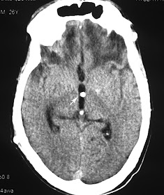
    Unusual migration of proximal tip of ventriculoperitoneal shunt - CT

    26 year old male patient with post traumatic hydrocephalus treated with ventriculoperitoneal shunt axial CT sectio at hte level of third ventricle showing tubular hyperdense structure (arrow) suggestive of VP shunt.

    Axial section at the level of basal cisterns showing the VP shunt catheter in the suprasellar structures.

    Sagittal reformatted image showing the catheter tip is situated at the roof of the sella.

    Discussion:
    • There are many incidents of unusual migration of the shunt tip in to various parts of the body. 
    • Usually it is the distal or the peritoneal catheter that migrates after breakage or disconnection into many locations such as the scalp, heart, anus/rectum, urethral, knee, umbilicus, chest, pleural cavity, inguinal canal and scrotum.
    • Complete migration of the shunt in to the ventricles have been reported.
    • This might be the first case of shunt migration in to the suprasellar region as far as my knowledge is concerned.

    Sunday, October 9, 2011

    Pottt's spine with bilateral psoas abscess - MRI


    Sagittal T2 and T1 images of MRI lumbar spine in 20 year old male showing partial collapse of the L4 vertebral body with restropulsion of the posterior fragment causing thecal sac compression and mild compression on the traversing nerve roots. 

    Axial T2 image at the level of L4 showing bilateral large psoas abscesses.
    Coronal image well depict the craniocaudal extent of the abscess.

    Lipoma Arborescens of the Knee - MRI

    Sagittal T1 weighted MRI in 27 year old male shows a large suprapatellar effusion with a frond-like synovial mass of fat intensity in suprapatellar region and in posterior intercondylar region (arrows).
    Sagittal PD FS MRI shows the synovial mass to be the same low intensity as fat (arrows).

    Axial gradient echo MRI shows the characteristic frond-like pattern.


    Discussion:
    Lipoma arborescens is a rare benign lesion, which arises in the synovium and is characterized by villous proliferation of fat cells. The proliferating cells appear organic and often resemble a tree thus its name, arborescens, from the Latin arbor for tree.

    Cause: is uncertain. One hypothesis is - synovial hyper-proliferation is in response to traumatic or inflammatory stimuli.
    Associations: Trauma, osteoarthritis or rheumatoid arthritis.

    Radiograph: Findings are non specific.
    1. Soft tissue density with areas of lucency suggesting fat.
    2. Underlying degenerative changes of the knee.

    MRI: is the investigation of choice.
    • Villous proliferation, often with a frond-like configuration, which arises from within the synovial cavity.
    • Fatty characteristics and typical pattern of proliferation shows isointense to the fat on all the sequences.
    • Joint effusion.
    • Meniscal  tears.
    • Baker's cyst in 38%.

    Intralobar sequestration - CT Angiography.

    31 year old male patient came with repeated respiratory tract infections. CT showed consolidation in right lower lobe. Mediastinal window images show suspicious small artery supplying the lesion from the descending thoracic aorta for which CT angio was done


    CT angio MIP and VRT images showing large single artery supplying the sequestrated segment of the lung. Venous drainage to the pulmonary veins. Later patient was taken for angioembolization of aberrant artery.

    Discussion:

    Definition: An aberrant lung tissue mass that has no normal connection with the bronchial tree or with the pulmonary arteries. 
    Arterial blood supply: Systemic arteries, usually the thoracic or abdominal aorta.
    Venous drainage: Azygous system, the pulmonary veins, or the inferior vena cava.

    Two types:  
    1. Intralobar sequestrations: can manifest as an area of increased opacity simulating pneumonia, as a mass with or without air-fluid levels, or as cysts. 
    2. Extralobar sequestration, in which the sequestered lung has its own separate pleural covering, is much less common and usually is found on the left side next to the hemidiaphragm. At radiography, it may manifest as a reasonably well-defined mass at the base of the left hemithorax. Rarely, an intralobar and extralobar sequestration may occur in the same patient.
    The main differentiating points are as given in the following table.


    Thursday, October 6, 2011

    Scapholunate ligament tear - MRI

    Radiograph of wrist in 45 Year old male patient with pain in the wrist joint showing widened scapholunate joint space with focal periosteal elevation in the lunate (arrow).

    Coronal PD FS image showing complete tear in the scapholunate ligament with mild marrow edema in the scaphoid and lunate bones.

    Coronal GRE image well depicts the ligament tear (arrow).
    Discussion:

    • Most common and most significant ligament injury of wrist causing carpal instability.
    • Risk factors: ulna minus configuation, slope of radial articular surface, and lunotriquetral coalition.
    Spectrum of injury include: (increasing severity)
    1. Dynamic scapholunate instability
    • No radiographic evidence of malalignment is present (ie dynamic deformity); 
    • Diagnosis is established by dorsal S-L tenderness and positive shift test; 
    2. Rotatory subluxation of scaphoid:
    3. Scapholunate dissociation (SLD):

    • SL ligament tear may lead to rotational dislocation of scaphoid allowing proximal pole to displace posteriorly & distal pole to displace anteriorly.
    • Scaphoid inherently tends to palmar flex because of its oblique position and the loading applied through (STT) joint.
    • Because scaphoid lacks proximal of ligament, it will rotate around radiocaptitate ligament leading to dorsal rotary subluxation of the proximal pole.
    4. Dorsal intercalated segment instability: (DISI) 
    5. Scapholunate advanced collapse:

    Osteosarcoma of femur - Radiograph and MRI

    Radiograph of 20 year old female showing destructive lytic lesion involving the metaphysis of the medial aspect of femur with new bone formation and periosteal reaction (caudman triangle) {Arrow}

    T1 weighted coronal image showing lytic destructive lesion with soft tissue, periosteal reaction and new bone formation.

    Axial GRE image showing the new bone formation as hypointense signal and periosteal reaction as hyperintense signals.

    Coronal STIR image showing the lesion.
    Discussion: Osteosacroma is malignant tumor of bone in which neoplastic osteoid is produced by a proliferating spindle cell stroma.

    • Most common primary malignant bone tumor of mesenchymal derivation.
    • Arises in adolescents, and second or third decade.
    • Affects males slightly more often than females. 

    Affected sites:
    • Occurs in region of knee (distal femur or proximal tibia) in 50% of patients. 
    • Other sites include proximal humerus, proximal femur, & pelvis.
    • Most osteosarcomas occur in the metaphysis.
    • Infrequently occurs in the spine.
    Classification:
    1. Classic 
    2. Telangiectatic 
    3. Parosteal 
    4. Periosteal
    IMAGING:
    Radiograph:
    • Destructive lytic lesion or mixture of lytic and sclerotic areas (common).
    • Moth eaten appearance with ill-defined zone of demarcation.
    • Involves the metaphysis
    • New bone formation.
    • Periosteal reaction
    • Visible soft-tissue mass.
    • Spiculated / Sunburst appearance.
    • 'Codman's Triangle' which is basically a subperiosteal lesion formed when the periosteum is raised due to the tumor.
    CT Scan:
    • Gives clearer indication of bone destruction.
    • May depict small amounts of mineralized osseous matrix not seen on radiographs. 
    • May be particularly helpful in visualizing flat bones, in which periosteal changes may be more difficult to appreciate.
    MRI:
    • Modality of choice in evaluating the local extent of disease because of its excellent bone marrow and soft tissue contrast and multiplanar capabilities.
    • it assists in determining the most appropriate surgical management.
    • Better delineates the involvement of the epiphysis, adjacent joint, adjacent soft tissues, neurovascular bundle, marrow extent and skip lesions.
    Bone Scans:
    • Increased uptake of radioisotope on bone scans obtained by use of technetium-99m (99m Tc) methylene diphosphonate (MDP).
    • most useful in excluding multifocal disease but skip lesions are more reliable on MRI.

    Wednesday, October 5, 2011

    Giant Cell Tumor of Fibular head - Radiograph and MRI.

    Radiograph of the knee in 32 year old male patient showing well defined expansile lytic lesion seen in the  epimetaphyseal region of fibular head with thinning of the cortex and cortical break at few places.

    Axial T2 FS image at the level of fibular head showing expansile lytic lesion showing fluid contents with septations, thinning of the cortex and cortical break at few places. No obvious blood fluid levels noted.

    Axial T1 weighted image at the level of fibular head showing the lesion with isointense contents and thinning of the cortex.

    Coronal T1 weighted images showing the lesion.
    Discussion:
    • GCT is a common benign but locally aggressive lesion of unknown etiology. 
    • It occurs chiefly in men between 20-50 yrs (after epiphyseal closure).
    • The tumor is expansile lytic lesion that involves the epiphysis & metaphysis.
    • The tumor may enlarge to occupy most of epiphysis & adjacent metaphysis. 
    • The tumor may erode & penetrate subchondral bone, articular cartilage, & cruciate ligaments.
    Location:

    • Epiphysis of distal femur, proximal tibia, & distal radius.
    • Other sites: fibula, sacrum, proximal humerus, & distal tibia.
    • Can occur in bones of pelvis, particularly ilium near SI joint and sacrum.
    Spine:
    • Usually located in vertebral body; 
    • Radiolucent lesion in vertebral body of a young patient is likely to be GCT; 
    Staging:
    Stage I:
    • Benign latent giant cell tumors.
    • No local aggressive activity.
    Stage II:
    • Benign active GCT. 
    • Imaging studies demonstrate alteration of the cortical bone structure.
    Stage III:
    • Locally aggressive tumors.
    • Imaging studies demonstrate a lytic lesion surrounding medullary and cortical bone.
    • There may be indication of tumor penetration through the cortex into the soft tissues.
    IMAGING:
    Radiographic Features: well-defined lytic lesion that involves the metaphysis and epiphysis (typical of a giant cell tumor);

    CT Scan: helps to determine the extact amount of cortical destruction and helps determine the optimal location of the cortical window;

    Bone Scans: Bone scans may show decreased radioisotope uptake in the center of lesion (doughnut sign). It is also found in ABC.

    MRI:
    • MRI May show fluid fluid or blood fluid levels.
    • Help determine determine extent of tumor destruction and soft tissue involvement.
    • May be indicated when the tumor has eroded through the cortex and allows determination of whether concomitant neurovascular structures are involved.
    • May help evaluate subchondral penetration.
    Differential diagnosis:
    1. Aneurysmal bone cyst - ABC.
    2. Non Ossifying fibroma.

    Wednesday, September 28, 2011

    Supraspinatus avulsion tear - MRI

    Proton density oblique coronal MR image in 41 year old male patient with trauma showing focal fracture in the greater tuberosity of the humerus (arrow head) with full thickness tear in the supraspinatus tendon and retraction of the tendon fibers (arrow) suggestive of full thickness avulsion tear.

    T1 TSE oblique coronal MR image showing focal fracture in the greater tuberosity of the humerus (arrow head) with absent hypointense  supraspinatus tendon.

    Axial T2 Medic (GRE) image showing fracture and tendon tear.

    Sagittal STIR image showing full thickness tear and absent tendon fibers.

    Discussion:
    • Full-thickness avulsion tears of the tendon away from the greater tuberosity are less common.
    • Massive tears often extend posteriorly to involve the infraspinatus tendon or extend anterior to tear, the anterior interval and subscapularis tendon.
    • If an acute complete supraspinatus tendon tear is identified, surgery is often scheduled within the next several days so that the tendon can be repaired before the development of retraction or atrophy.
    • Angled oblique sagittal T2/STIR images perpendicular to the plane of the supraspinatus tendon can better delineate the partial and full thickness tears.

    Tuesday, September 27, 2011

    Calvarial thickening - MRI

    T1 weighted axial image in 24 year old female with thalasemia showing diffuse thickening of the calvarium.

    T2 weighted axial image showing calvarial thickening.

    FLAIR axial image showing calvarial thickening.

    Discussion:
    Differential diagnosis of calvarial thickening:
    1. Various severe anemias - bone marrow expansion.
    2. Pagets disease.
    3. Hyperparathyroidism.
    4. Osteopetrosis.
    5. Chronic Dilatin therapy.
    6. Acromegaly.
    7. Some rare genetic diseases - Camurati-Engelmann’s disease, frontometaphyseal dysplasia and   craniodiaphysial dysplasia.

    Traumatic optic nerve compression - CT

    Axial CT section through the optic nerves in a 59 year old male with trauma showing fracture in the posterior part of the lateral wall of the right orbit and the fracture fragment is causing compression on the optic nerve at the optic canal (arrow) and note the normal wide optic canal on left side (arrow head).

    Coronal CT scan bone window showing the bony fragment compressing the optic nerve and narrowing the optic foramen (arrow) and note the normal foramen on left side (arrow head).

    Sagittal image showing the bony fragment compressing on the optic nerve.
    Discussion:
    • Traumatic optic neuropathy is a devastating potential complication of closed head injury.
    • The hallmark of an optic neuropathy, traumatic or otherwise, is a loss of visual function, which can manifest by subnormal visual acuity, visual field loss, or color vision dysfunction.
    • Vision loss associated with traumatic optic neuropathy can be partial or complete and temporary or permanent.
    • Best diagnostic test is Thin-slice CT scan of the nose, sinuses, and orbits
    • Fractures through the optic canal can be best depicted with thin-section CT scanning (eg, 1.5-mm cuts with 1-mm intervals).
    • CT scanning provides adequate imaging of orbital soft tissue and is better than MRI at delineating bony defects. 
    • It provides an intraoperative road map for the surgeon in patients who require surgical decompression and can be used for image guided endoscopic surgery.
    • Treatment is observation, steroids and optic nerve decompression. The decision for surgical decompression should still be based primarily on the clinical examination findings.

    Cystosarcoma phyllodes tumor

    Film screen mammogram of the right breast in 41 year old female showing large well defined dense lesion with smooth margins. No calcification. 


    USG of the same patient showing heteroechoic mass lesion with well defined smooth margins. The lesion shows width>breadth. No evidence of calcification.

    Discussion:
    Cystosarcoma phylloides is a rare breast tumor that accounts for approximately 0.5% of breast tumors examined at biopsy and approximately 2.5% of-fibroid epithelial tumors examined at biopsy. This tumor was named in 1838 by Johannes Muller, who described large firm tumors of the breast. He believed the tumor to be benign and observed that the surface had a leaf-like (phylloide) configuration and contained narrow cyst-like spaces within a sarcoma- like stroma.

    It was not until 1943 that Cooper and Ackerman reported on the malignant biological potential of this tumor. Clinically, patients present with a palpable breast mass. Although periods of rapid growth are known to occur, the mass usually develops slowly. The lesion is mobile, not fixed to the skin or the chest wall. Although the lesions have occasionally been noted in both breasts, they are usually found only in one. They have been reported to occur in patients of any age, although the average age is 40 to 45 years.

    Mammogram usually shows well defined mass lesion of varying sizes, predominantly large mass. The lesion shows smooth margins in benign and irregular margins in malignant lesion. There may or may not have macrocalcification. USG shows solid mass lesion within the breast and shows a defined margins and some small fluid filled clefts with in the lesion may be diagnostic of cystosarcoma phylloides.

    Sunday, September 11, 2011

    Usual Interstitial Pneumonia with Tuberculosis - HRCT

    Radiograph (scanogram)  of 81 year old male with dyspnea showing reticular opacities, fibrotic bands, honey coombing and few fibrocavitatory lesions in bilateral upper lobes consistent with UIP with old Tuberculosis.


    HRCT of 81 Year old female showing macrocystic honeycombing and reticular
    opacities predominantly in the subpleural region.

    HRCT along the upper lobes showing fibrocavitatory lesions

    Coronal lung window showing reticular shadows and honey combing predominantly in the basal region with fibrocavitatory lesion in upper lobes.

    Usual Interstitial Pneumonia - HRCT

    HRCT of 40 Year old female showing macrocystic honeycombing, reticular
    opacities predominantly in the subpleural region, architectural distortion and
    areas of focal ground-glass opacity 

    Mediastinal window of the same patient showing small enlarged pre tracheal lymphnode.

    Discussion:

    Idiopathic Pulmonary Fibrosis (IPF) is the most common entity of the Idiopathic Interstitial Pneumonias (IIPs). By definition, IPF is the term for the clinical syndrome associated with the morphologic pattern of UIP.
    Median survival - 2 to 4 years.

    Clinical features - Presents in 50 years old or older. Patients present with progressively worsening
    dyspnea and nonproductive cough.

    Histopathologic Features: Spatial and temporal heterogeneity, architectural distortion and fibroblastic foci.

    HRCT: Distribution is apicobasal gradient and predomonently subpleural location. 
    Features are macrocystic honeycombing, reticular opacities, traction bronchiectasis, architectural distortion and focal ground-glass opacity.