Tuesday, April 26, 2011

Renal angiomyolipoma-CT

Axial CT of kidneys in a 29 year old male showing small exophytic fat attenuation lesion in the left kidney (arrow) suggestive of angiomyolipoma.

Zoomed picture of the above showed image better shows the lesion.

Discussion:

Angiomyolipoma (AML) is the most common benign tumour of the kidney and is composed of blood vessels, smooth muscle cells and fat cells. It is strongly associated with tuberous sclerosis.

Imaging features:
Ultrasonography (USG) - Well defined hyper echoic lesion in the kidney. Hyperechogenecity is due to fat content of the lesion.

CT Scan: Well defined fat attenuation lesion as shown in our case. CT angiography may be helpful to identify the aneurysms which predict the fatal hemorrhage.

MRI - Hyperintense on both T1 and T2 weighted images due to its fat content and appears hypointense on fat suppressed T1 images. 
If the fatty tissue is scanty in-phase and out-phase images T1 weighted sequence is very helpful in identifying the fat component in the lesion, which is seen as loss of signal on out-phase images.
People with tuberous sclerosis needs yearly follow up  renal scans.
Treatment:
  • If the lesion is < 4 cm - follow up imaging to look for the progression of the lesion.
  • If it is > 4 cm or presence of aneurysm, needs to be treated with trans arterial embolisation or surgical excision. Embolising agents used are PVA (Polyvinyl alcohol) and absolute alcohol mixed with lipiodol. 

Renal cell carcinoma with Hiatus hernia-CT

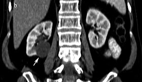
Axial CT of kidneys in a 42 year old male showing large heterogeneously enhancing lesion in the left kidney with perinephric extension.

Axial section at the level of diaphragm of the same patient showing hiatus hernia (arrow) 

Duplex collecting system with ureterocele- CT Urography.


3D volume rendering maximum intensity projection image of CT urography in 29 year old male showing duplication of collecting system, upper and mid ureter on the left side (arrow head) with orthotopic ureterocele (arrow)

Sunday, April 24, 2011

Duplex collecting system-CT Urography.


3D volume rendering maximum intensity projection image of CT Urography in 27 year old male showing duplication of collecting system, upper and mid ureter (arrow). Both the ureters are joining in their lower thirds (arrow head).

Papillary necrosis - CT Urography

Maximum intensity projection image of CT Urography in 32 year old male showing papillary necrosis in the right kidney with calyx cut off (arrow) due to old Koch’s

Renal parenchymal scarring-CT Urography


Coronal reformatted CT Urography images in a 32 year old male showing cortical scarring in bilateral kidneys due previous tuberculosis

Renal calculus-NCCT KUB (Stone protocol)


Axial and coronal reformatted non contrast CT images in a 32 year old female showing left renal calculus (arrows).

Renal cyst - CT


Axial and coronal reformatted CT images in a 36 year old male showing simple cortical cyst in the right kidney (Bosniak category I cyst).

Ureteric calculus-NCCT KUB (Stone protocol)


Axial and coronal reformatted non contrast CT images in a 41 year old showing right ureterovesical junction calculus (arrows).
Non contrast CT in multi slice CT (stone protocol) is the modality of choice for the diagnosis of ureteric calculus.

Fibromuscular dysplasia-CT Renal Angiography.


Volume rendered CT angiography image in 25 year old male scheduled for donor nephrectomy showing irregularity (string of beads) in the bilateral renal arteries (arrows) more so on the right side with aneurysm (arrow head) in one of the distal lobar renal artery branch.

Renal artery stenosis


Thick oblique axial and oblique coronal maximum intensity projection CT images in a 48 year old male donor scheduled for laparoscopic nephrectomy showed renal artery stenosis at the origin on the left side (arrow) with calcific focus.

Late confluence of left renal vein with Large gonadal vein draining the left renal vein


Thick oblique coronal maximum intensity projection CT image in a 46 year old female donor scheduled for laparoscopic nephrectomy showed late confluence of the left renal vein tributaries with large left gonadal vein (arrow) draining inferior tributary of renal vein (arrowhead). 
It is very important to identify this gonadal vein to prevent complication during laparoscopic nephrectomy.

Renal vein variants- Duplicated left inferior vena cava

Thick oblique sagittal maximum intensity projection CT images in a 26 year old female donor scheduled for donor nephrectomy showed duplication of inferior venacava (arrow).

Renal vein variants-Double renal vein

Thick maximum intensity projection CT image in a 41 year old female donor scheduled for donor nephrectomy showing double renal vein on the right side (arrows). Incidentally note multiple calcified gall stones (arrow head).

Renal vein variants-Retroaortic vein


Axial and thick maximum intensity projection CT images in a 46 year old female donor scheduled for donor nephrectomy showing retroaortic left renal vein (arrows) and double renal vein on the right side (arrow head).

Discussion:
Most common is multiple renal veins, seen in approx 15-30% of individuals. 
The most common anomaly of the left renal venous system is the circumaortic renal vein, seen in up to 17% of patients 
I. There are two common variants of the circumaortic vein: 
1. The most common variant (approximately 75% of cases), one renal vein at the renal hilum subsequently divides before entering the inferior vena cava. 
2. The less common variant, two distinct veins originate from the renal hilum.

II. A less common venous anomaly is the complete retroaortic renal vein, seen in 3% of patients. Here, the single left renal vein courses posterior to the aorta and drains into the lower lumbar portion of the inferior venacava.
Alternatively, the retroaortic renal vein can drain into the iliac vein.

Renal artery variants- High origin of renal artery

Volume rendered CT angiography image in a 26 year old male donor scheduled for donor nephrectomy showing abnormally high origin (D11 level) of the right renal artery. It is important to indentify the level of origin for the operating surgeon.
Volume rendered CT angiography image in another person aged 46 year old scheduled for donor nephrectomy showing abnormally high origin (D12 level) of the right renal artery.

Renal artery variants- early branching

Volume rendered CT angiography images in a 34 year old male donor scheduled for donor nephrectomy showing early branching (<2 cm) on the right side (arrow).

Discussion:
Branching of the renal artery at a distance of less than 1.5/2 cm from its origin is early branching.

A large early branch from the renal artery can result in complications during the laparoscopic donor nephrectomy and during the renal transplantation.

Thursday, April 21, 2011

Renal artery variants- Accessory renal artery


3D Volume rendered maximum intensity projection (MIP) CT angio images in a 29 year old male donor scheduled for donor nephrectomy showing Accessory renal artery on the left side with prehilar branching on right side.

Discussion:
  • Multiple renal arteries are unilateral in approximately 30% of patients and bilateral in approximately 10%. 
  • Accessory arteries usually arise from the aorta or iliac arteries anywhere from the level of T11 to the level of L4. 
  • In rare cases, they can arise from the lower thoracic aorta or from lumbar or mesenteric arteries. 
  • Usually, the accessory artery courses into the renal hilum to perfuse the upper or lower renal poles. 
  • Prehilar arterial branching is another common variant that must be checked in patients being evaluated for donor nephrectomy.

Thursday, April 14, 2011

CT Renal angiography - Normal

 3D volume rendering image showing normal bilateral renal arteries and aorta
Coronal curved multi planar reformatted image showing normal bilateral renal veins.

Renal arteries: 
  • Single renal arteries bilaterally that originates from the abdominal aorta at the level of L3 below the origin of superior mesenteric artery. 
  • It is important to identify the variants of the renal arteries.

Renal veins:
  • Usually lies anterior to the renal artery at the renal hilum. 
  • Left is 3 times longer than right. 
  • Left – 6-10 cm length- courses anterior to aorta. 
  • Right – 2-4 cm in length. The left renal vein receives left gonadal, left adrenal and left lumbar tributaries.

Perianal Fistula - Grade 1

Coronal STIR image of the 48 year old male patient showing linier hyperintense tract ascending superiorly in the perianal area and curving back medially and inferiorly (arrow).

T2 Fat suppressed image showing the horizontal portion of the fistulous tract (arrow).

2 Fat suppressed image showing the termination of the fistula in the inter-sphincteric region suggestive of grade I perianal fistula.

Discussion and another case of grade I perianal fistulahttp://manju-imagingxpert.blogspot.com/2011/04/perianal-fistula-grade-1.html


Monday, April 11, 2011

Spondylolisthesis-MRI

T2 weighted sagittal image of the lumbar spine in a 44 year old male patient showing grade I spondylolisthesis at L3-4 (anterolisthesis of L3 over L4) with disc herniation. No traversing nerve roots compression. 

T2 weighted sagittal image of the lumbar spine right lateral most image and the left lateral most images showing exiting nerve root compression (arrow) and break in the pars inter articularis suggesting spondylolysis.

Axial T2 weighted image at L3-4 showing nerve root compression bilaterally.

Discussion:

Spondylolisthesis is officially categorized into five different types by the Wiltse classification system: Dysplastic, Isthmic, Degenerative, Traumatic, and Pathologic.

Grades:
  • Grade 1 is 0–25%
  • Grade 2 is 25–50%
  • Grade 3 is 50–75%
  • Grade 4 is 75–100%
  • Over 100% is Spondyloptosis, when the vertebra completely falls off the supporting vertebra.

Disc extrusion - MRI


T2 weighted sagittal MR images of the lumbar spine showing central disc extrusion with cranial migration.

Discussion:
A disk is classified as an extrusion if any distance between the edges of the disk material beyond the disk space is greater than the distance between the edges of the base measured in the same plane.

Extruded disk material that has no continuity with the disk of origin may be further characterized as sequestrated. A sequestrated disk is a subtype of extruded disk. By definition, a sequestrated disk can never be classified as a protruded disk. Disk material that is displaced away from the site of extrusion, regardless of the presence or absence of continuity, may be called migrated, a term that is useful for interpreting images because it is often impossible to show continuity on imaging.

Friday, April 8, 2011

Perianal Fistula - Grade 1

T2 weighted image showing well defined linier hyperintense tract in the 12 o' clock position of the perianal region terminating in the inter sphincteric plane (arrow) suggestive of grade 1 simple inter sphincteric fistula. No ramification or abscess formation.
T1 weighted axial image showing the fistula (tract)

Visit the following link for MR Imaging Classification of Perianal Fistulas and Its Implications for Patient Management

Perianal Fistula - Grade 3


T2 weighted image showing well defined linier hyperintense tract in the 12 o' clock position of the perianal region terminating in the trans sphincteric plane (arrow) suggestive of grade 3 trans sphincteric fistula. No ramification or abscess formation.


T2 Fat saturated sagittal image showing the tract (arrow)

Discussion:
Perianal fistulization is an uncommon but important condition of the gastrointestinal tract that causes substantial morbidity. Perianal fistulas occur in approximately 10 of 100,000 persons, with a twofold to fourfold male predominance. Magnetic resonance (MR) imaging has been shown to demonstrate accurately the perianal anatomy.

Grading:

St James's University Hospital MR Imaging Classification of Perianal Fistulas
Grade
Description
0
Normal appearance
1
Simple linear intersphincteric fistula
2
Intersphincteric fistula with intersphincteric abscess or secondary fistulous track
3
Trans–sphincteric fistula
4
Trans–sphincteric fistula with abscess or secondary track within the ischioanal or ischiorectal fossa
5
Supralevator and translevator disease


Epidermoid

T2 weighted axial image of the 22 year old gentle man showing well defined hyperintense mass lesion in the basal cisterns on right side causing significant mass effect on the temporal horn of ipsilateral lateral ventricle.

T1 weighted axial image showing the lesion is hypointense to gray matter.


FLAIR axial image the lesion is showing mixed signal intensity predominantly hypointense.

Coronal T2 WI showing the lesion.

Axial B1000 Diffusion weighted image showing moderate restriction.

ADC image showing hypointense mass, which differentiates epidermoid from arachnoid cyst where the lesion will be hyperintense on ADC images.

Discussion:

Epidermoid cysts (sebaceous cysts) are benign congenital lesions of ectodermal origin. They account for approximately 1% of all intracranial tumors. Although these lesions are congenital, patients are usually not symptomatic until they are aged 20-40 years.

CT scan:

  • Low attenuation - similar to CSF density.
  • No enhancement.
  • Calcification in 15-20%.
  • Rarely they can be hyperdense secondary to high density.
MRI:
  • Isointense to CSF on T1 and T2 weighted images. T1 signal intensity varies with the amount of lipid content.
  • May be hyperintense on FLAIR images.
  • High signal on diffusion weighted images which differentiate from the low signal arachnoid cyst.
  • Minimal rim enhancement on post contrast images.

Arachnoid cyst
  • CSF filled/signal
  • Middle cranial fossa (MC) > cerebral convexities, basal cisterns, retrocerebellar region
  • Hypogenesis of adjacent brain
  • No enhancement
  • Low on DWI (like ventricle).

Friday, April 1, 2011

Frontoethmoidal Encephalocele

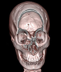
13 Year old female with hard swelling in the fore head. Volume rendering colour coded image of the face showing fromtoethmoidal encephalocele.

Volume rending image with bone algorithm showing bony prominence in the frontoethmoidal region. 

Cropped volume rendered image to show the defect in the frontoethmoid region

Oblique coronal reformatted bone window image showing the defect. 

Sagittal reformatted bone window image showing the defect clearly.

Coronal T2 fat saturated MR image showing herniation of the meninges through the above showed defect.

Discussion:

Encephalocele can be a congenital or an acquired abnormality of the brain in which intracranial contents including meninges, CSF, and brain tissue herniate through a skull defect. Congenital encephaloceles occur when the mesodermal layer between the neural tube and the ectoderm fails to develop and the anterior neuropore remains open. Anterior encephaloceles can also occur after trauma or surgery. Brain pulsations are presumed to push brain tissue through the defect. Patients are prone to recurrent episodes of meningitis. In addition, visual acuity and hypothalamic function may be affected. Clinical presentation includes a nasopharyngeal mass, which may enlarge with Valsalva's maneuver.

Differential considerations include a tumor traversing the cribriform plate, granuloma or esthesioneuroblastoma.